Nano News
Looking inside Nanolive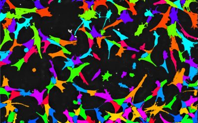
Watch Nanolive’s new webinar: “Label-free analysis of living cell populations reveals controlled phenotypic variation among single cells”
Nanolive is delighted to announce that our webinar with the topic "Label-free analysis of living cell populations reveals controlled phenotypic variation among single cells" is now available on demand. Watch it here. In this webinar, Dr. Emma Gibbin, Communications...
Nanolive label-free live cell imaging weighs in on the fight against antibiotics resistance
Table of content Multi-drug resistant (MDR) bacteria are a potent threat to public health Bacteriophages offer a source of hope for solving the antibiotics resistance crisis Endolysins can be native or engineered, but how can they be administered? Testing the...
Competition – 6 months free usage of Nanolive’s Automated Live Cell Imaging Solution
Call-out to all pharma and biotech companies To conclude this very “special” year with an optimistic outlook into the future, we call all pharma and biotech companies to tell us how you would use live cell imaging in your research, to help solve one of the “Ten...
5 new scientific publications using Nanolive label-free live cell imaging
We are delighted to announce 1, 2, 3, 4, 5 exciting new articles featuring Nanolive cell imaging. The articles come from users based in Poland, Germany, and the USA. The in vitro toxicity evaluation of halloysite nanotubes (HNTs) in human lung cells by Sawicka et...
Nanolive receives Microscopy Today Innovation Award 2020 for automated live cell imaging microscope CX-A
Nanolive are delighted to have been selected as winners of the 11th annual Microscopy Today Innovation Awards in 2020 for the development of our automated microscope, the CX-A, a non-invasive live cell imaging method for continuous organelle monitoring in cell...

Watch Nanolive’s new webinar “Unlocking the mysteries of neurite growth in primary cortical neurones: a quantitative approach to live cell imaging”
We are happy to announce that our webinar with the topic "Unlocking the mysteries of neurite growth in primary cortical neurones: a quantitative approach to live cell imaging" is now available on demand. In this webinar, Dr. Emma Gibbin, Communications Specialist at...
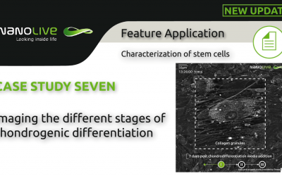
Imaging label-free live chondrogenic differentiation, with a focus on the early morphological changes mesenchymal stem cells undergo on the way to becoming cartilage tissue
Background Chondrogenic differentiation describes how cartilage is formed from mesenchymal stem cells (MSCs). Cells pass two morphological stages; first they differentiate into chondroblasts, which then mature into chondrocytes. Chondroblasts are responsible for the...
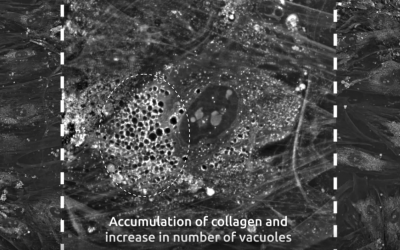
Chondrogenic differentiation of mesenchymal stem cells imaged live, using Nanolive’s label-free, non-invasive imaging
Chondrogenic differentiation: from stem cell to cartilage Mesenchymal stem cells (MSCs) are multipotent stromal cells that can differentiate into a variety of cell types including osteoblasts (bone cells), neurones (nerve cells), chondrocytes (cartilage cells),...
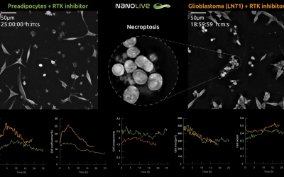
How do live cells respond to receptor tyrosine kinase (RTK) inhibition?
Glioblastoma: the most aggressive brain cancer Glioblastoma multiforme (GBM) is an aggressive brain tumor that places among the most lethal of cancers. Unfortunately, most patients with GBMs die of their disease in less than a year and long-term survival prospects are...
Do you want to image stress fibers – the architecture of the cell – for unlimited periods of time at high frequency?
Stress fibers: what are they and why are they important? Stress fibers form the architecture of the cell. They are made of bundles of filaments composed from two strands of actin that are bound together by cross-linking proteins (see Figure 1). Their alignment,...
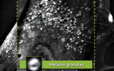
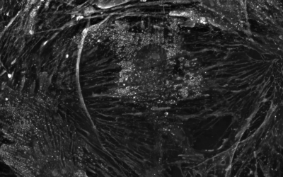
Non-invasive, label-free visualization of melanin granules using Nanolive cell imaging
What is the function of melanin? Melanin describes the group of natural pigments that is responsible for skin and hair pigmentation and photoprotection of the skin and eye. Deficiency in melanin can cause several disorders and diseases including vitiligo, deafness,...
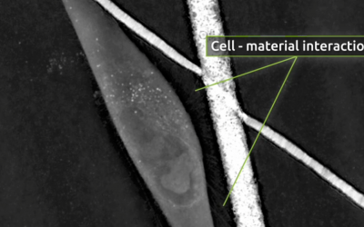
Is surface roughness the key to improving bone scaffolds?
We are delighted to announce a new publication featuring the 3D Cell Explorer, has been published in the scientific journal, Materials & Design. The study, led by Prof. Urszula Stachewicz at the AGH University of Science and Technology in Kraków, Poland, used...
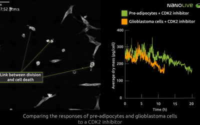
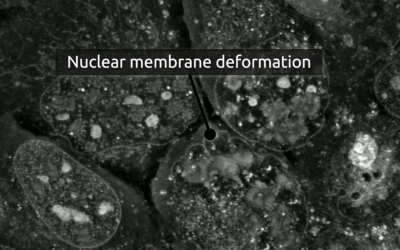
High-resolution time-lapse imaging of cell response to CDK2-inhibition
One of the major advantages of Nanolive’s label-free imaging is that images can be acquired at high frequency, over long periods of time. This allows us to analyse the responses of cells to drug-perturbation with high temporal precision. Here, we observe how...
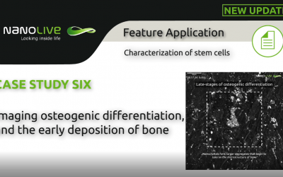
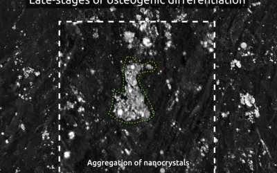
Imaging osteogenic differentiation, and the early deposition of bone
Background Osteogenesis describes the process of bone formation. The cells that create new layers of bone tissue are called osteoblasts. These cells are differentiated from mesenchymal stem cells (MSCs), in a linear sequence. The dynamics of the morphological and...
4 new scientific publications using Nanolive cell imaging
We are delighted to announce FOUR exciting new articles featuring Nanolive live cell imaging. The articles come from users based in Poland, Switzerland, and Australia. Apoptosis as the main type of cell death induced by calcium electroporation in...
Is it possible to capture horizontal mitochondrial transfer in real time using Nanolive cell imaging?
Mitochondrial transfer Studies have demonstrated that mitochondria and mitochondrial DNA can be transferred between cells, but the function of this, and the mechanisms involved in the transfer remain highly debated [1]. Some studies have shown that mitochondrial...
Capturing the deposition and early growth of bone that occur during osteogenic differentiation
Osteogenesis is the process in which new layers of bone tissue are placed by osteoblast cells, capable of synthesizing and mineralization bone. Osteoblasts are differentiated from mesenchymal stem cells (MSCs), in a linear sequence progressing from osteoprogenitors...
Morphological changes during adenovirus infection in osteosarcoma cells
In this footage, obtained using Nanolive’s 3D Cell Explorer-fluo we observe the progression of GFP-labelled adenovirus in human bone osteosarcoma epithelial (U2OS) cells. Adenovirus is known to migrate to the nucleus of its host cell where it forms structures known as...