Stress fibers: what are they and why are they important?
Stress fibers form the architecture of the cell. They are made of bundles of filaments composed from two strands of actin that are bound together by cross-linking proteins (see Figure 1). Their alignment, distribution and shape play a vital role in cell, adhesion, motility, and mechanosensing and they are fundamental to almost all biological processes.
Co-localization of fluorescence stain with refractive index maps shows label-free visualization of stress fibers is possible
Here, we stained mesenchymal stem cells with SiR-actin, a highly specific fluorescent probe for F-actin, and captured both fluorescent images (Cy5 filter) and refractive index (RI) images simultaneously, using the 3D Cell Explorer-fluo.
Images were taken every minute for 10 hours. After, RI maps were overlaid with the fluorescent images to visualize where the stress fibers are located.
In the video, we show the fluorescence images intermittently, to make it easier to colocalize the images with the RI maps.
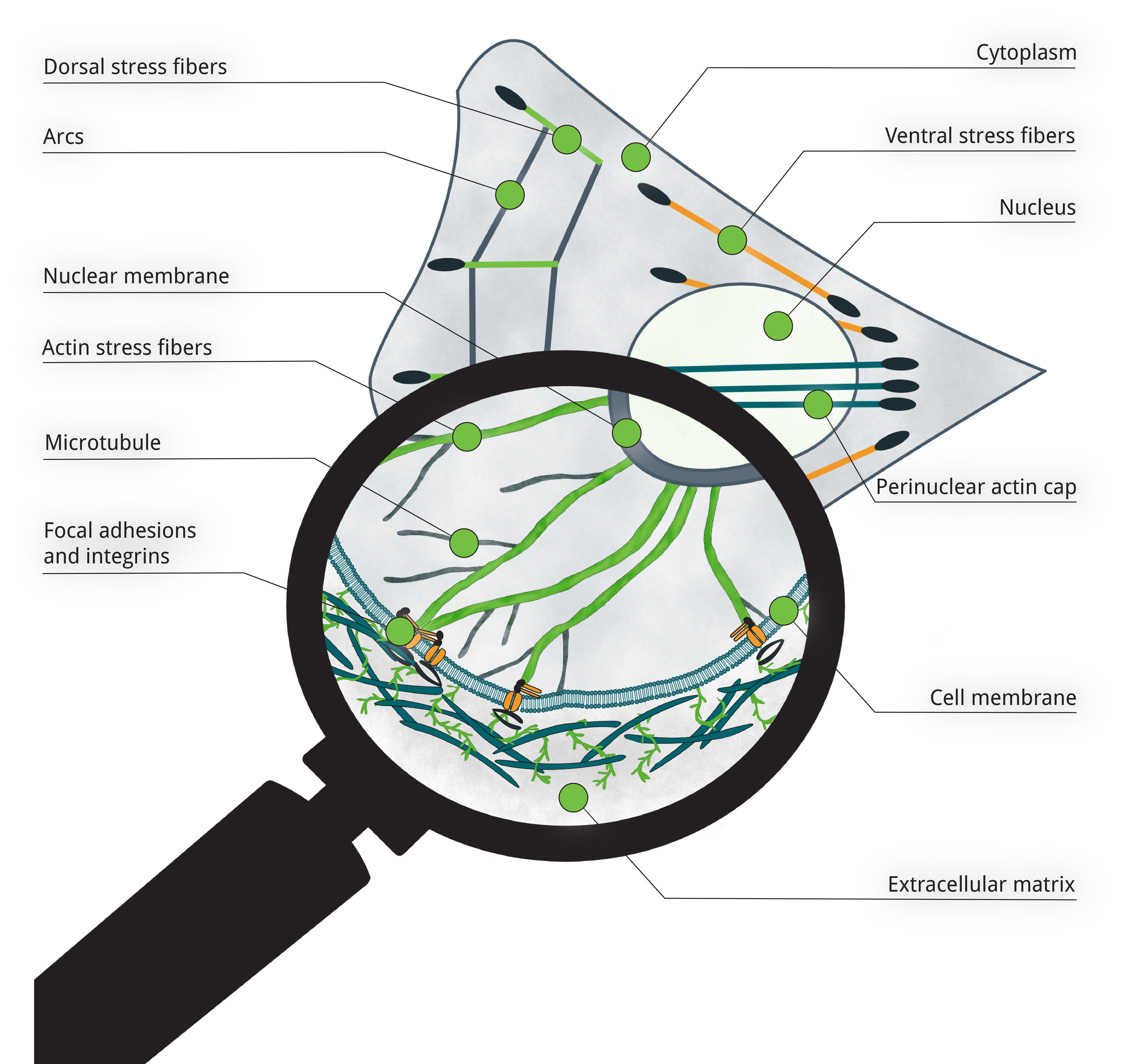
Figure 1
Stress fibers: distribution in the cell, and a closer look on their structure. Schema adapted from Burridge, K., & Guilluy, C. (2016). Focal adhesions, stress fibers and mechanical tension. Experimental cell research, 343(1), 14–20 and Wang, N., “Infographic: Following the Force, Physical forces propagate from the outside of cells inward and vice versa”, The Scientist, 31 Jan. 2017, https://www.the-scientist.com/infographics/infographic-following-the-force-32114.
What are the pros and cons of fluorescence-based imaging?
This beautiful footage highlights the ubiquity and importance of stress fibers.
Fluorescence is an important proof-of-concept that it is possible to capture stress fibers label-free using Nanolive imaging, but it also imposes restrictions on how long cells can be imaged for. Phototoxicity quickly becomes a problem; damaging samples and eventually, causing cell death.
By the end of the video, we start to see bubbles being formed. These are clear signs of cellular stress, which can be avoided by using Nanolive cell imaging alone.
What does Nanolive cell imaging have to offer for future studies of stress fibers?
With Nanolive cell imaging researchers can monitor cells for long periods of time. This can be leveraged to study how stress fibers re-model the actin cytoskeleton, in control conditions or in response to drug-perturbation. From a biological perspective the applications of Nanolive label-free imaging are endless. They could be used to investigate (for example) the dynamic formation, arrangement, and recycling of stress fibers that facilitate cell migration, or the role that stress fibers play in cell division, endocytosis, and cell migration.
If you are interested in exploring some of these possibilities, then contact a member of our team today! Just click here.
Read our latest news

Cytotoxic Drug Development Application Note
Discover how Nanolive’s LIVE Cytotoxicity Assay transforms cytotoxic drug development through high-resolution, label-free quantification of cell health and death. Our application note explores how this advanced technology enables real-time monitoring of cell death...

Investigative Toxicology Application Note
Our groundbreaking approach offers a label-free, high-content imaging solution that transforms the way cellular health, death, and phenotypic responses are monitored and quantified. Unlike traditional cytotoxicity assays, Nanolive’s technology bypasses the limitations...

Phenotypic Cell Health and Stress Application Note
Discover the advanced capabilities of Nanolive’s LIVE Cytotoxicity Assay in an application note. This document presents a detailed exploration of how our innovative, label-free technology enables researchers to monitor phenotypic changes and detect cell stress...
Nanolive microscopes

3D CELL EXPLORER
Budget-friendly, easy-to-use, compact solution for high quality non-invasive 4D live cell imaging

3D CELL EXPLORER-fluo
Multimodal Complete Solution: combine high quality non-invasive 4D live cell imaging with fluorescence

CX-A
Automated live cell imaging: a unique walk-away solution for long-term live cell imaging of single cells and cell populations