Nanolive's Imaging Technology
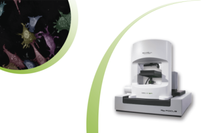
Real-time, label-free and high resolution images of living cells Contact usNanolive imaging platforms use holotomography to capture label-free timelapse images of cells and their organelles to give insight into dynamic processes and phenotypic changes, but how does the technology work? This video explains the concept of holotomography, and the unique technology behind Nanolive imaging.
Video: What is holotomography? This video explains the concept of holotomography, and the unique technology behind Nanolive imaging
What is holotomography?
Holography
Imaging objects quantitatively using the phase-shift of light
+
Tomography
The process of assembling information from cross sections to achieve 3D imaging
Video: Long Term Live Imaging of Mouse Embryonic Stem Cells imaged with Nanolive
Combining these two methods enables high resolution imaging of cells and their organelles in 3D, making this a high-content imaging technique which is well-suited for fundamental in vitro research and drug development. Furthermore, this label-free method is non-invasive and free of both phototoxicity, and harmful effects of chemical dyes. This means cells can be imaged for days without harm, enabling investigation of long-term processes like stem cell differentiation.
What is Nanolive’s unique approach to holotomography?
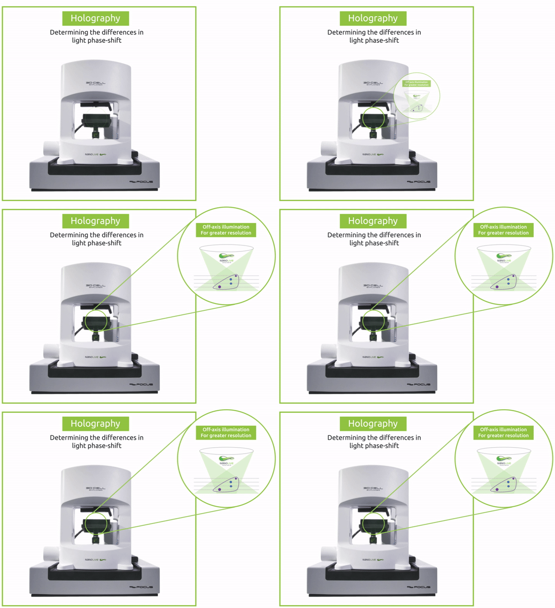
- Holography uses variations in the phase-shift of light as it passes through an object to produce quantitative images.
- As low-power light passes through a sample, the wave is altered by the properties of the sample’s internal structure.
- Cellular structures and organelles interact with light in different ways, meaning they can be differentiated by their refractive index values.
- This structural information is encoded on a detector by comparing the object beam with a reference beam of light. The phase-shift between the two light beams is retrieved through computation based on the recorded hologram.
How does Nanolive’s holotomography achieve such high resolution?
- In order to achieve a higher resolution than is possible using phase-shift imaging alone, Nanolive systems are equipped with a patented rotating arm which rotates the object beam 360° around the sample.
- 100 snapshots are taken from different angles to achieve a resolution of 200nm in the x, y.
How does tomographic imaging reveal intracellular landscapes?
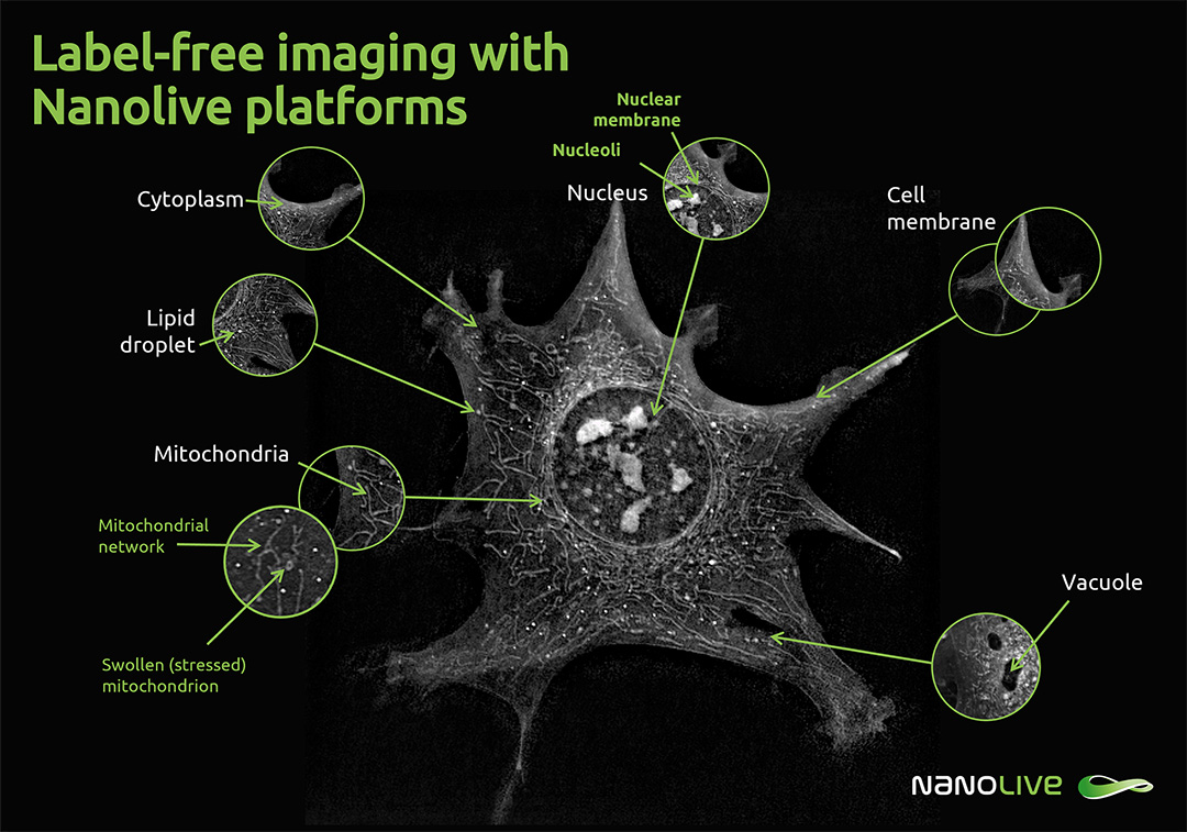
96 2D image slices are taken in the z-stack, covering 30µm in depth, allowing internal organelles like nucleoli and mitochondria to be visualized clearly.
What does a holotomogram look like?
Download the guide to label-free organelle identification here.
- By combining all the images, a high-resolution 3D holotomographic reconstruction is produced, which can be represented as a 2D maximum intensity projection as you see on the image here.
- In this image, refractive index is indicated by brightness; darker areas like the cell medium and cytoplasm are darker, as the refractive index is lower. Brighter areas like the small dots which are lipid droplets have a higher refractive index.
- This allows quantification of the dry mass of cells and their organelles.
- Holotomographic images provide insights into 3D processes of cells and their organelles, like the sub-cellular localisation and organisation of organelle systems, membrane dynamics, cell-cell interactions, identifying the mechanism of cell death, and many other phenotypic changes.
How does Nanolive utilize Holotomography and AI to unlock cellular insights?
These high-resolution, quantitative images, are rich in data, which can be extracted by AI analysis. Nanolive provides a suite of digital analysis solutions to automate this process and to quantify cell death, effector cell killing, lipid droplet dynamics, and other unique phenotypic changes.
Learn more about Nanolive’s Imaging Technology with the miniseries “Discover Label-free Imaging Technology With Nanolive”

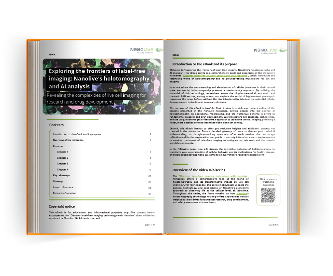
Nanolive’s technology eBook
“Exploring the frontiers of label-free imaging: Nanolive’s holotomography and AI analysis”
Revealing the complexities of live cell imaging for research and drug development
This eBook serves as a comprehensive guide and expansion on the innovative miniseries “Discover label-free imaging technology with Nanolive”, which introduces the fascinating world of holotomography and its groundbreaking implications for live-cell imaging.

Contact us and a Nanolive expert will get in touch with you to discuss your needs.


