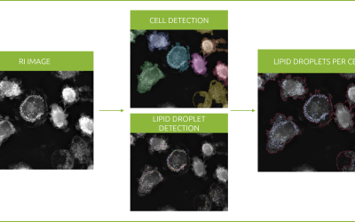
Nanolive are delighted to have been selected as winners of the 11th annual Microscopy Today Innovation Awards in 2020 for the development of our automated microscope, the CX-A, a non-invasive live cell imaging method for continuous organelle monitoring in cell populations and 96-well plates. The award is shared with 10 other top technologies that have high potential to advance microscopy in the areas of light microscopy, electron microscopy and microanalysis.
This is the second award Nanolive have won for the CX-A, which was also awarded The Scientist Top 10 Innovations in 2019.
We would like to extend our congratulations to Chief Technology Officer Dr. Sebastien Equis and acknowledge the important contributions of Dr. Alexandre E. Grandchamp, Julien Renggli and Philippe Paccaud throughout the development process. Thanks also go to the whole R&D team and their dedication and hard work to achieve this high quality product.
Read our latest news
Revolutionizing lipid droplet analysis: insights from Nanolive’s Smart Lipid Droplet Assay Application Note
Introducing the Smart Lipid Droplet Assay: A breakthrough in label-free lipid droplet analysis Discover the power of Nanolive's Smart Lipid Droplet Assay (SLDA), the first smart digital assay to provide a push-button solution for analyzing lipid droplet dynamics,...
Food additives and gut health: new research from the University of Sydney
The team of Professor Wojciech Chrzanowski in the Sydney Pharmacy School at the University of Sydney have published their findings on the toxic effect of titanium nanoparticles found in food. The paper “Impact of nano-titanium dioxide extracted from food products on...
2023 scientific publications roundup
2023 has been a record year for clients using the Nanolive system in their scientific publications. The number of peer-reviewed publications has continued to increase, and there has been a real growth in groups publishing pre-prints to give a preview of their work....
Nanolive microscopes
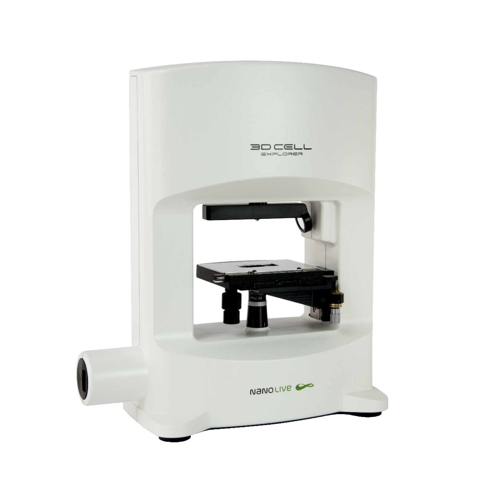
3D CELL EXPLORER
Budget-friendly, easy-to-use, compact solution for high quality non-invasive 4D live cell imaging
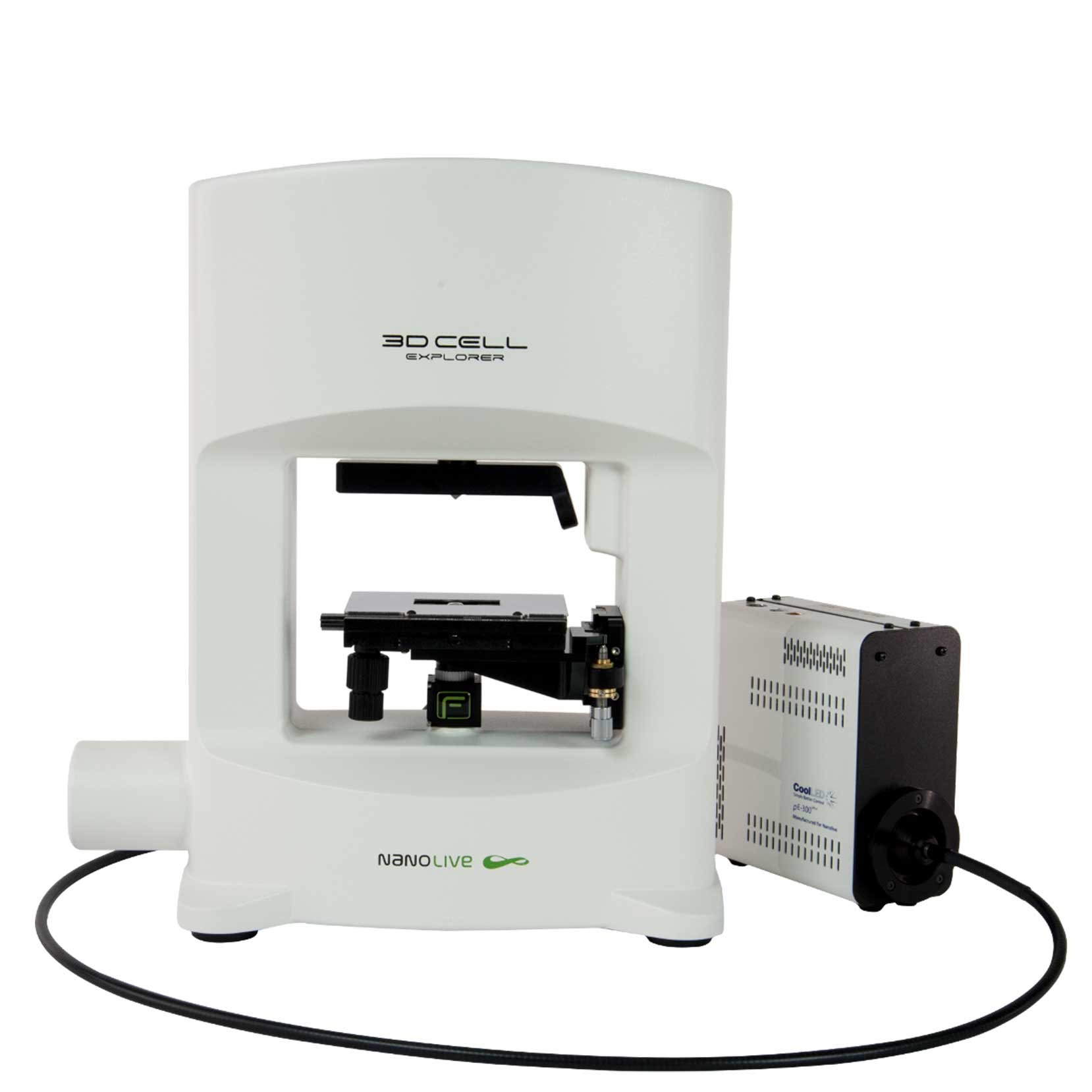
3D CELL EXPLORER-fluo
Multimodal Complete Solution: combine high quality non-invasive 4D live cell imaging with fluorescence
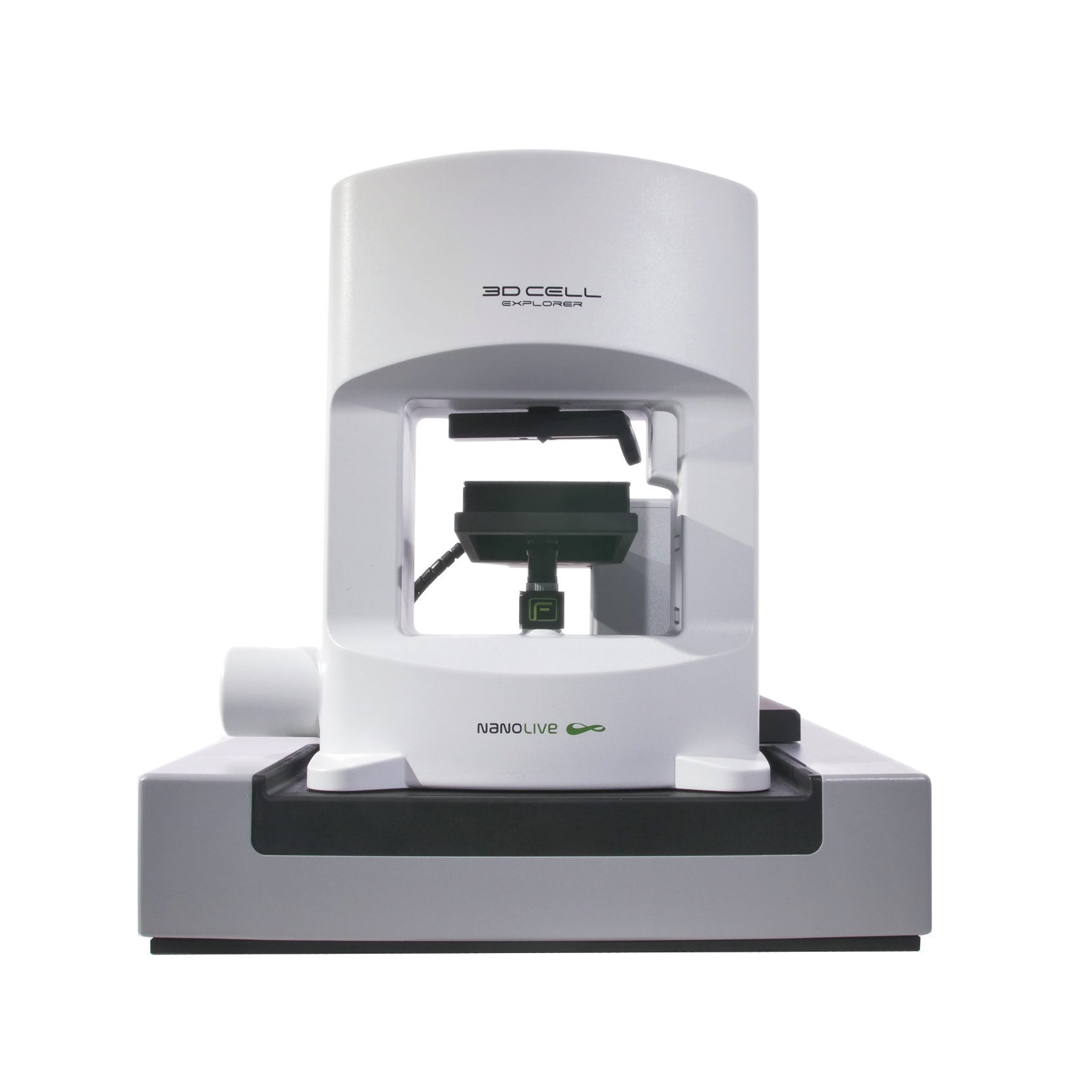
CX-A
Automated live cell imaging: a unique walk-away solution for long-term live cell imaging of single cells and cell populations

