Mitochondrial transfer
Studies have demonstrated that mitochondria and mitochondrial DNA can be transferred between cells, but the function of this, and the mechanisms involved in the transfer remain highly debated [1].
Some studies have shown that mitochondrial transfer can restore the function of defective mitochondria in recipient cells [2,3]. While other studies have demonstrated that it plays a role stem cell differentiation [4,5], and mediating inflammatory.
The structures/mechanisms involved in mitochondrial transfer are just as diverse. Tunneling nanotubes, microvesicles, mitochondrial ejection and cytoplasmic fusion are just a few of the hypotheses generated to explain mitochondrial transmission [7].
Using Nanolive live cell imaging for mitochondrial transfer observations
In this video (one image taken every 2 mins), we appear to capture intercellular transfer of mitochondria in non-perturbed pre-adipocyte cells. Our footage suggests that initial contact between cells occurs via long thin cellular extensions and that subsequent interactions involve the broad cytoplasmic projections of both cells.
If this is the case, Nanolive cell imaging may be able to shed light on the mechanisms involved in mitochondrial transfer.
Bibliography
[1] Paliwal S, Chaudhuri R, Agrawal A, Mohanty S. 2018. Regenerative abilities of mesenchymal stem cells through mitochondrial transfer. J. Biomed. Sci. 25(1):1-2.
[2] Spees JL, Olson SD, Whitney MJ, Prockop DJ. 2006. Mitochondrial transfer between cells can rescue aerobic respiration. Proc. Natl. Acad. Sci. 103(5):1283-8.
[3] Lin HY, Liou CW, Chen SD, Hsu TY, Chuang JH, Wang PW, Huang ST, Tiao MM, Chen JB, Lin TK, Chuang YC. 2015. Mitochondrial transfer from Wharton’s jelly-derived mesenchymal stem cells to mitochondria-defective cells recaptures impaired mitochondrial function. Mitochondrion. 22:31-44.
[4] Vallabhaneni KC, Haller H, Dumler I. 2012. Vascular smooth muscle cells initiate proliferation of mesenchymal stem cells by mitochondrial transfer via tunnelling nanotubes. Stem Cells Dev. 21(17):3104-13.
[5] Acquistapace A, Bru T, Lesault PF, Figeac F, Coudert AE, Le Coz O, Christov C, Baudin X, Auber F, Yiou R, Dubois‐Randé JL. 2011. Human mesenchymal stem cells reprogram adult cardiomyocytes toward a progenitor‐like state through partial cell fusion and mitochondria transfer. Stem Cells. 29(5):812-24.
[6] Court AC, Le‐Gatt A, Luz‐Crawford P, Parra E, Aliaga‐Tobar V, Bátiz LF, Contreras RA, Ortúzar MI, Kurte M, Elizondo‐Vega R, Maracaja‐Coutinho V. 2020. Mitochondrial transfer from MSCs to T cells induces Treg differentiation and restricts inflammatory response. EMBO Rep. 5;21(2):e48052.
[7] Torralba D, Baixauli F, Sánchez-Madrid F. 2016. Mitochondria know no boundaries: mechanisms and functions of intercellular mitochondrial transfer. Front. Cell Dev. Biol. 28;4:107.
Read our latest news
Cytotoxic Drug Development Application Note
Discover how Nanolive’s LIVE Cytotoxicity Assay transforms cytotoxic drug development through high-resolution, label-free quantification of cell health and death. Our application note explores how this advanced technology enables real-time monitoring of cell death...
Investigative Toxicology Application Note
Our groundbreaking approach offers a label-free, high-content imaging solution that transforms the way cellular health, death, and phenotypic responses are monitored and quantified. Unlike traditional cytotoxicity assays, Nanolive’s technology bypasses the limitations...
Phenotypic Cell Health and Stress Application Note
Discover the advanced capabilities of Nanolive’s LIVE Cytotoxicity Assay in an application note. This document presents a detailed exploration of how our innovative, label-free technology enables researchers to monitor phenotypic changes and detect cell stress...
Nanolive microscopes

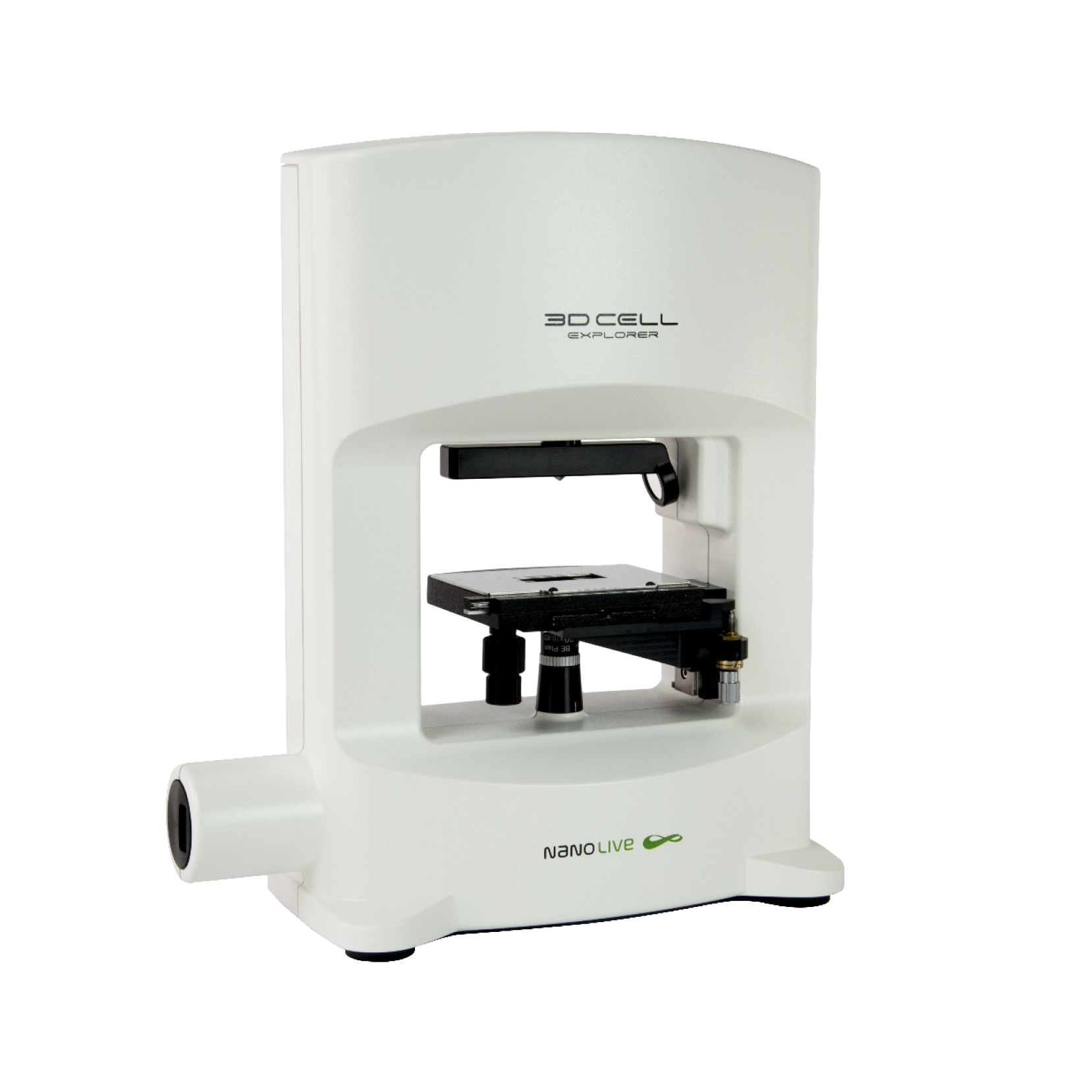
3D CELL EXPLORER
Budget-friendly, easy-to-use, compact solution for high quality non-invasive 4D live cell imaging

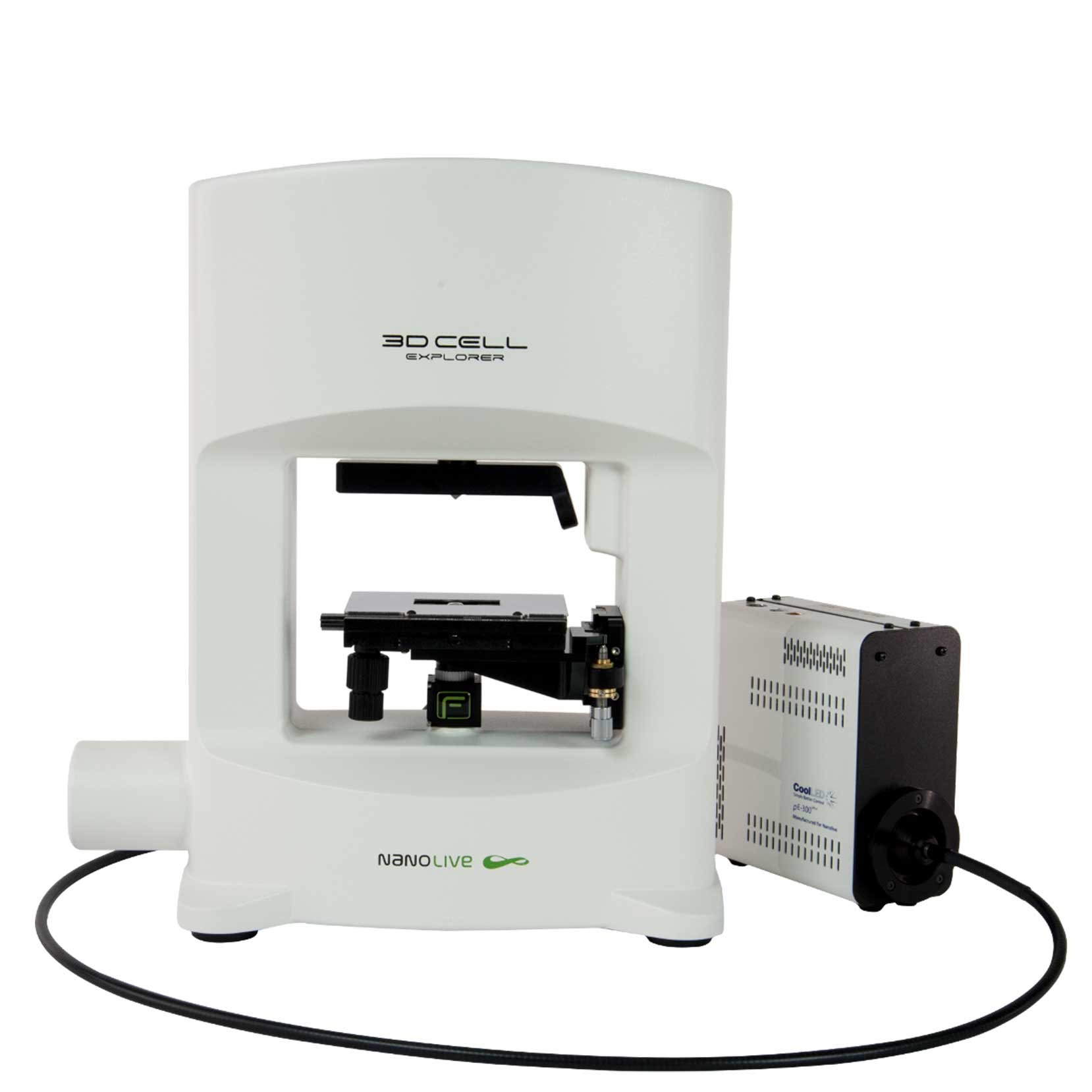
3D CELL EXPLORER-fluo
Multimodal Complete Solution: combine high quality non-invasive 4D live cell imaging with fluorescence

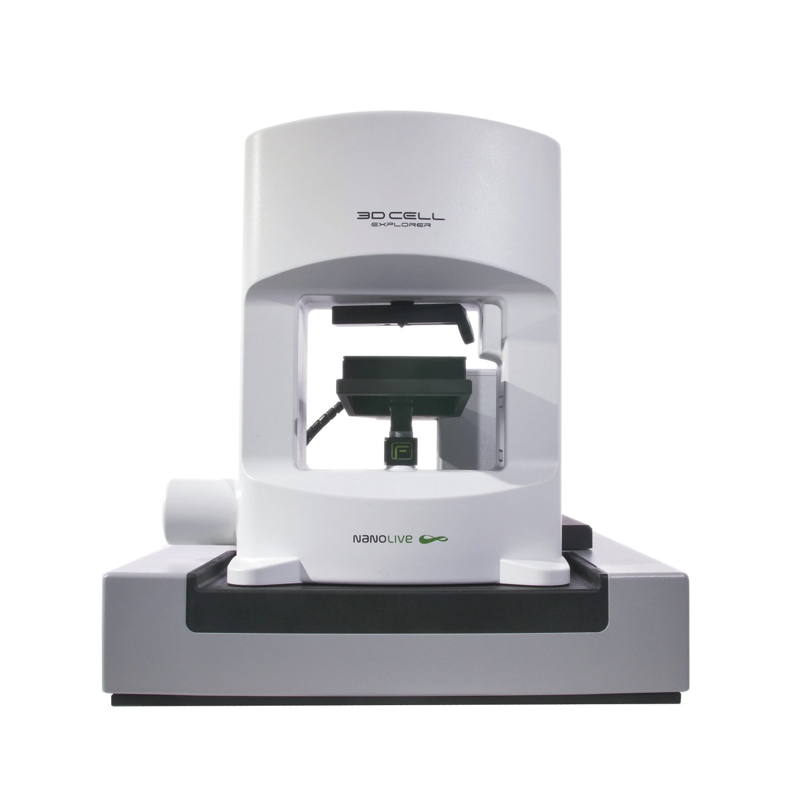
CX-A
Automated live cell imaging: a unique walk-away solution for long-term live cell imaging of single cells and cell populations


