Oncology & Immuno-oncology
Long-term non-invasive live cell imaging of the interactions between immune and cancer cells with Nanolive Request a demo or quoteNanolive technology allows to visualize the changes in cell morphology and behaviour that occur during interactions between immune cells and cancer cells at all stages of the disease process, from invasion, migration and metastasis, to cell death. Quantitative analyses of cell proliferation across time can also be performed enabling researchers to compare how cancer cells respond to different antibodies or immune checkpoint inhibitors.
New digital assay: The LIVE T Cell Assay is the first multi-parametric, label-free live immuno-oncology assay that is able to measure and characterize in one experiment how T cells find, bind, stress, kill and serial kill their targets and to select the best T cell therapy on living cells.
“The Nanolive technology is like putting on 3D glasses in a cinema – visualizing in depth cellular phenomena that are otherwise opaque and obscure.”
Prof. Douglas Hanahan
LIVE T Cell Assay
Label-free AI-assisted segmentation of target cells and T cells in co-culture
The LIVE T Cell Assay offers a unique opportunity to understand T cell/target cells interactions over time, at both the single-cell and population level and completely label-free. Nanolive’s label-free live cell imaging captures detailed, multiplexed, and texturally rich images of the entire T cell response process: find, bind, stress and kill.
Only Nanolive’s rich images can achieve a clear segmentation of T cells and their targets without using any destructive labels.
Testing the efficacy of bispecific antibodies
Cytotoxic T-cells are powerful effector cells capable of killing target cells bearing an appropriate antigenic complex (peptide–MHC), which is recognized by their T-cell receptor (TCR). Most T-cells are however unable to recognize and kill tumor cells, because of the lack of tumor-specificity of their TCR. By using bispecific antibodies targeting tumor-associated antigens (TAA) on the cancer cells and the CD3 protein complexes on T-cells, it is possible to redirect and activate any circulating T-cells against tumor cells independently of the specificity of their TCR.
In this video, we follow this process, using the 5×5 grid scan mode on Nanolive’s automated microscope, the CX-A. Image were acquired ever 4 min 30 s, for 20 h.
Read our detailed blogpost here.
Quantifying cell responses to Cdk2-inhibition
Cyclin-dependent kinase 2 (Cdk2) drives the progression of cells into the S- and M-phases of the cell cycle. The upregulation of Cdk2 activity plays a critical role in the growth of tumors in multiple cancers, and so the development of selective Cdk2 inhibitors hold promise for anticancer drug development.
In this video, we compare the dry mass changes that pre-adipocyte (green) and glioblastoma cells (orange) undergo in response to Cdk2 inhibition. Cells were imaged using the 5×5 gridscan mode (450×450 µm) on the CX-A, at an acquisition rate of 1 image every 2 secs for 20 h.
Both cell types lost dry mass following Cdk2 inhibition and as expected, cell death occurred during mitosis.
Read our detailed blogpost here.
Identifying different types of cell death
Receptor tyrosine kinases (RTKs) are key regulatory signaling proteins involved in cancer cell growth and metastasis, and so, they are attractive therapeutic targets for the treatment of cancer. In this video, we expose pre-adipocyte (green) and glioblastoma cells (orange) to an RTK-inhibitor and imaged their response. One image was acquired every 113 secs using the 5×5 gridscan mode on Nanolive’s CX-A.
RTK-inhibition initiated different mechanisms of cell death in the two cell types. Pre-adipocyte cell death occurred predominantly by apoptosis, whereas glioblastoma cell death occurred primarily by necroptosis. Understanding the type of cell death that a drug causes is important as it can affect the progression of diseases or the side-effects of a drug: apoptosis is immunologically silent meaning it does not promote a significant auto-immune response, whereas necroptosis induces significant inflammation.
Read the full blogpost here.
Combining label-free imaging with correlative epifluorescence
Chromatin stability play a key role in degenerative processes such as aging and cancer. This video, taken by Dr. Caroline Mauvezin, a Research Associate based at Bellvitge Biomedical Research Institute – IDIBELL in Barcelona (Spain) illustrates how Nanolive imaging can be used in conjunction with fluorescence (here green localizes Histone 2B-GFP a chromosome marker, and red, LAMP1-RFP a lysosome marker), to investigate such phenomena. Refractive index images were taken every 2 mins, and fluorescent images were captured once every 20 mins, for 5 h.
For more inspirational videos from researchers based in Spain and Portugal, check out our dedicated blogpost here.
Scientific Publications
Nanolive label-free live cell imaging has already shed light on many important topics in the field of immuno-oncology and oncology research. To get inspired and learn how your research can benefit from our technology, we invite you to check out these scientific articles published by our clients.
Webinar
Watch our Webinar: Explore detailed use-case data from our key opinion leaders, analysed by the LIVE Cytotoxicity Assay & LIVE T Cell Assay
Watch our Webinar: Accelerating oncology drug discovery with label-free imaging
In this webinar, we present the following use-cases of our immuno-oncology and cytotoxicity assays from industry-leading biopharma companies to test toxicity, selectivity, uptake and expression of different targeted therapies.
Specifically:
- Immune cell therapeutics:
- Bispecific antibody screening in a T cell-target cell co-culture (Lightchain Bioscience)
- Drug selectivity and cytotoxicity:
- mRNA expression and cytotoxicity (TrON)
- Drug selectivity in co-cultures: senolytics (dsm-firmenich)
- Measuring particle uptake (bit.bio)
In this webinar, Dr. Emma Gibbin-Lameira, Communications Specialist at Nanolive will discuss the advantages of using label-free live cell imaging in the drug discovery process and show how Nanolive’s automated live cell imaging solution the CX-A, can be used to:
- observe unique drug-induced phenotypes that fluorescence microscopy cannot capture
- record dynamic cellular responses to drug perturbations
- automate in vitro screening studies.
Dr. Gibbin-Lameira will present novel results from two experimental screens designed to test the phenotypic and morphological responses of pre-adipocyte and cancer cells to 8 inhibitors or modulators of kinase activity.
Please register here to view the webinar on-demand:
Please register here to view the webinar on-demand:
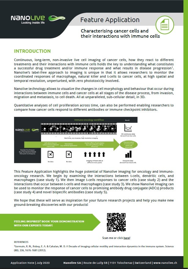
Feature Application
Characterising cancer cells and their interactions with immune cells
One of the most promising avenues of research in immune oncology is long-term, live imaging. Capturing dynamic biological processes in real time holds the key to understanding which cell-cell interactions promote a successful immune response, and which promote disease.
Video library
Activated T-cell killing a cancer cell
Read the blogpost here
Bispecific antibody induces T-cell proliferation and tumor cell killing
Read the blogpost here
T-cell attack and cancer evasion
Human peripheral blood mononuclear cells hunt melanoma cells
How can Nanolive imaging contribute to immuno-oncology research?
Read the blogpost here
HeLa cells are resistant to 8 nM TNF-α
Chloroquine-induced apoptosis in HeLa cells
Antibody Drug Conjugate (ADCs) killing mouse breast cancer cells
Read the blogpost here
Restored phagocytosis of cancer cells.
Read the blogpost here
Nanolive imaging and analysis platforms
Swiss high-precision imaging and analysis platforms that look instantly inside label-free living cells in 3D

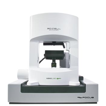
3D CELL EXPLORER 96focus
Automated label-free live cell imaging and analysis solution: a unique walk-away solution for long-term live cell imaging of single cells and cell populations

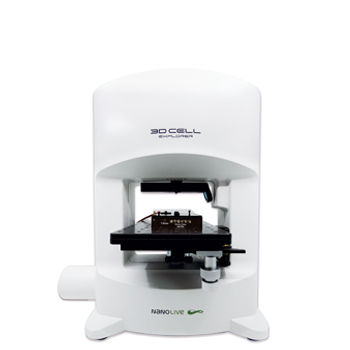
3D CELL EXPLORER-fluo
Multimodal Complete Solution: combine high quality non-invasive 4D live cell imaging with fluorescence

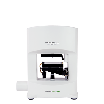
3D CELL EXPLORER
Budget-friendly, easy-to-use, compact solution for high quality non-invasive 4D live cell imaging

