Osteogenesis is the process in which new layers of bone tissue are placed by osteoblast cells, capable of synthesizing and mineralization bone. Osteoblasts are differentiated from mesenchymal stem cells (MSCs), in a linear sequence progressing from osteoprogenitors cells to pre-osteoblasts, osteoblasts, and finally, osteocytes [1].
Progression between the different stages is regulated by various transcription factors, cytokines, growth factors and extracellular matrix (ECM) proteins, that induce several important morphological changes [2].
Here, we use Nanolive’s CX-A to capture the morphological and physiological changes that occur during osteogenic differentiation. The label-free nature of our technology allowed us to image the process from day 1 to day 13, in a non-perturbed state, and at high temporal resolution (1 image taken every 10 mins using the 5×5 gridscan mode). MSCs were grown on fibronectin-coated dishes until 100% confluency was reached and then MSC osteogenic differentiation medium (C-28013, PromoCell) was added.
In the early differentiation stages, cells display the characteristic fibroblast-like phenotype of MSCs (i.e. cells are large, flat, and spindle-shaped). Some cells are motile and continue to divide by mitosis. Small, dynamic bright white spots can be observed near (or sometimes within) cells. The high refractive index (RI) of these structures suggest the material is dense; these could be calcium phosphate nanocrystals, which are deposited on the ECM.
After a one week in the osteogenic differentiation medium, cell division ceases and motility is reduced. Some (but not all) cells have started to become rounder; a classic sign that differentiation is underway [3]. There is also an increase in the number and size of the individual nanocrystals.
By the end of the experiment, static crystal aggregates can be observed. This is the first-time osteogenic differentiation and bone deposition has been captured label-free, in real time. We hope that this technique can shed light on 1) the osteogenesis process and 2) the pathogenesis of common bone diseases such as rickets, osteomalacia, osteoporosis and osteogenesis.
[1] Rutkovskiy A, Stensløkken KO, Vaage IJ. 2016. Osteoblast differentiation at a glance. Med. Sci. Monit. 22:95.
[2] Long MW, Robinson JA, Ashcraft EA, Mann KG. 1995. Regulation of human bone marrow-derived osteoprogenitor cells by osteogenic growth factors. J. Clin. Invest. 1;95(2):881-7.
[3] Pablo Rodríguez J, González M, Ríos S, Cambiazo V. 2004. Cytoskeletal organization of human mesenchymal stem cells (MSC) changes during their osteogenic differentiation. J. Cell Biochem. 1;93(4):721-31.
Read our latest news
Cytotoxic Drug Development Application Note
Discover how Nanolive’s LIVE Cytotoxicity Assay transforms cytotoxic drug development through high-resolution, label-free quantification of cell health and death. Our application note explores how this advanced technology enables real-time monitoring of cell death...
Investigative Toxicology Application Note
Our groundbreaking approach offers a label-free, high-content imaging solution that transforms the way cellular health, death, and phenotypic responses are monitored and quantified. Unlike traditional cytotoxicity assays, Nanolive’s technology bypasses the limitations...
Phenotypic Cell Health and Stress Application Note
Discover the advanced capabilities of Nanolive’s LIVE Cytotoxicity Assay in an application note. This document presents a detailed exploration of how our innovative, label-free technology enables researchers to monitor phenotypic changes and detect cell stress...
Nanolive microscopes

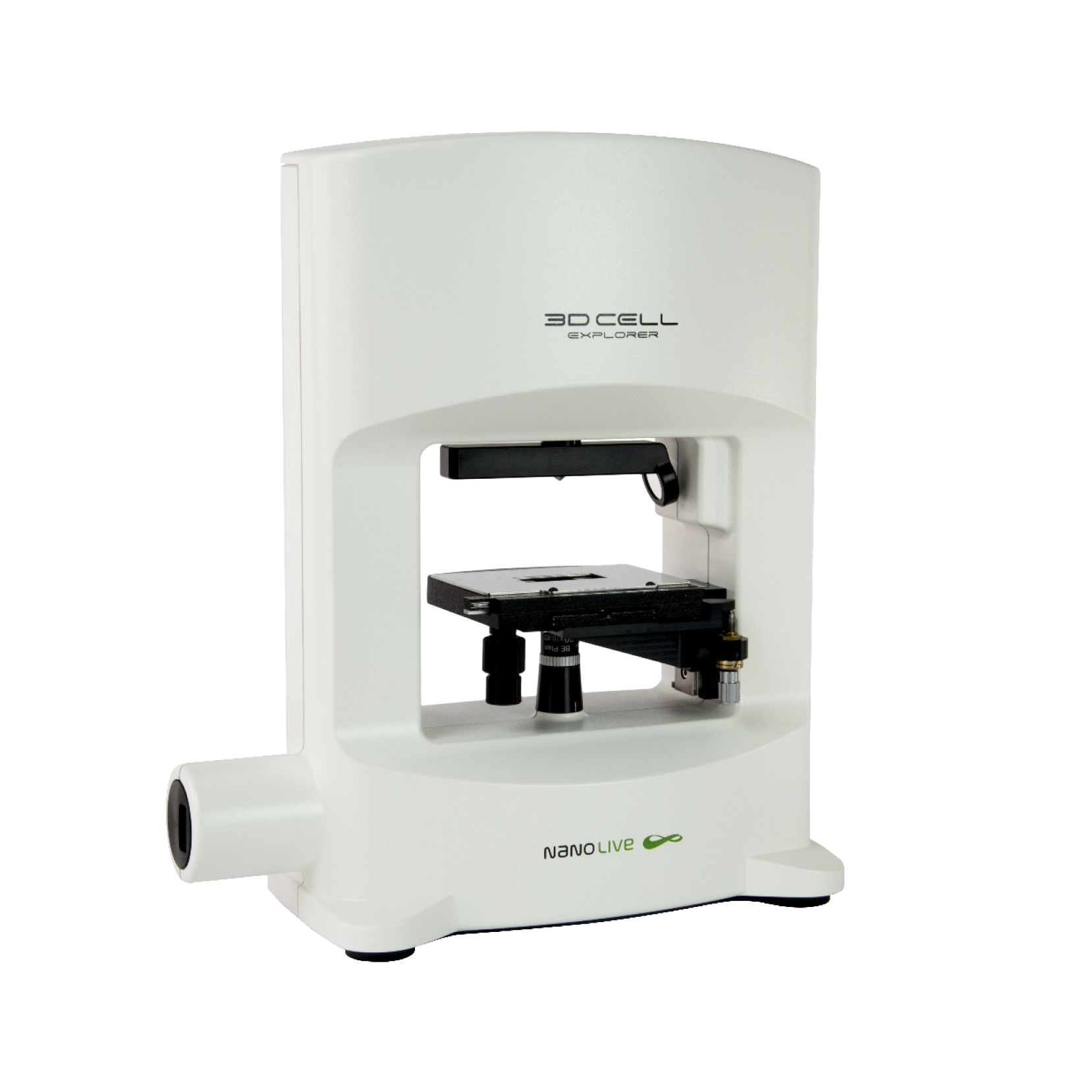
3D CELL EXPLORER
Budget-friendly, easy-to-use, compact solution for high quality non-invasive 4D live cell imaging

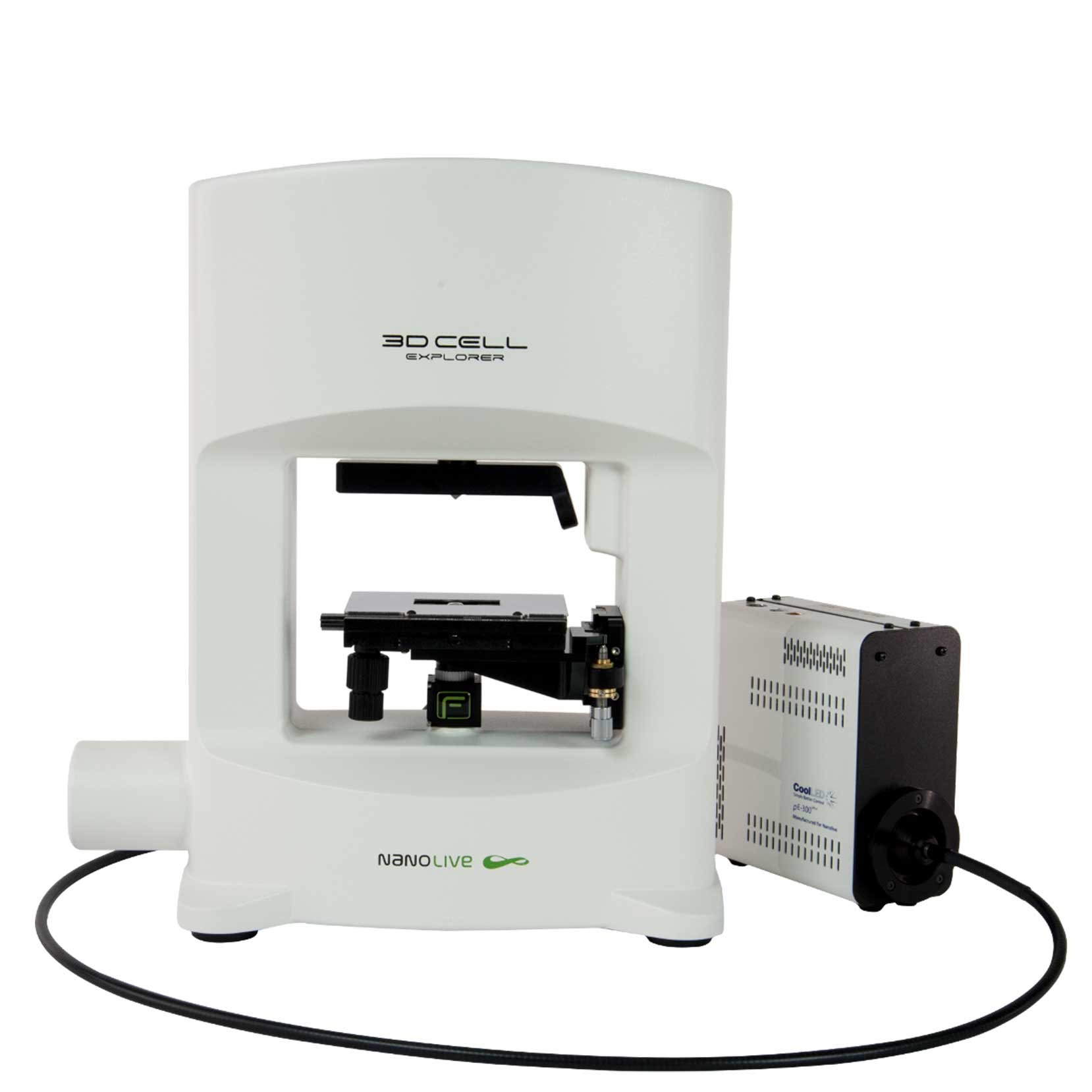
3D CELL EXPLORER-fluo
Multimodal Complete Solution: combine high quality non-invasive 4D live cell imaging with fluorescence

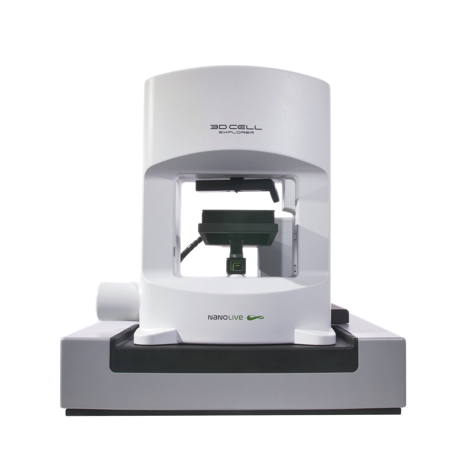
CX-A
Automated live cell imaging: a unique walk-away solution for long-term live cell imaging of single cells and cell populations


