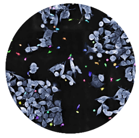
The 3D Cell Explorer 96focus
Bringing unlimited high content analysis to label-free live cell imaging EnquireGet higher significance earlier and faster
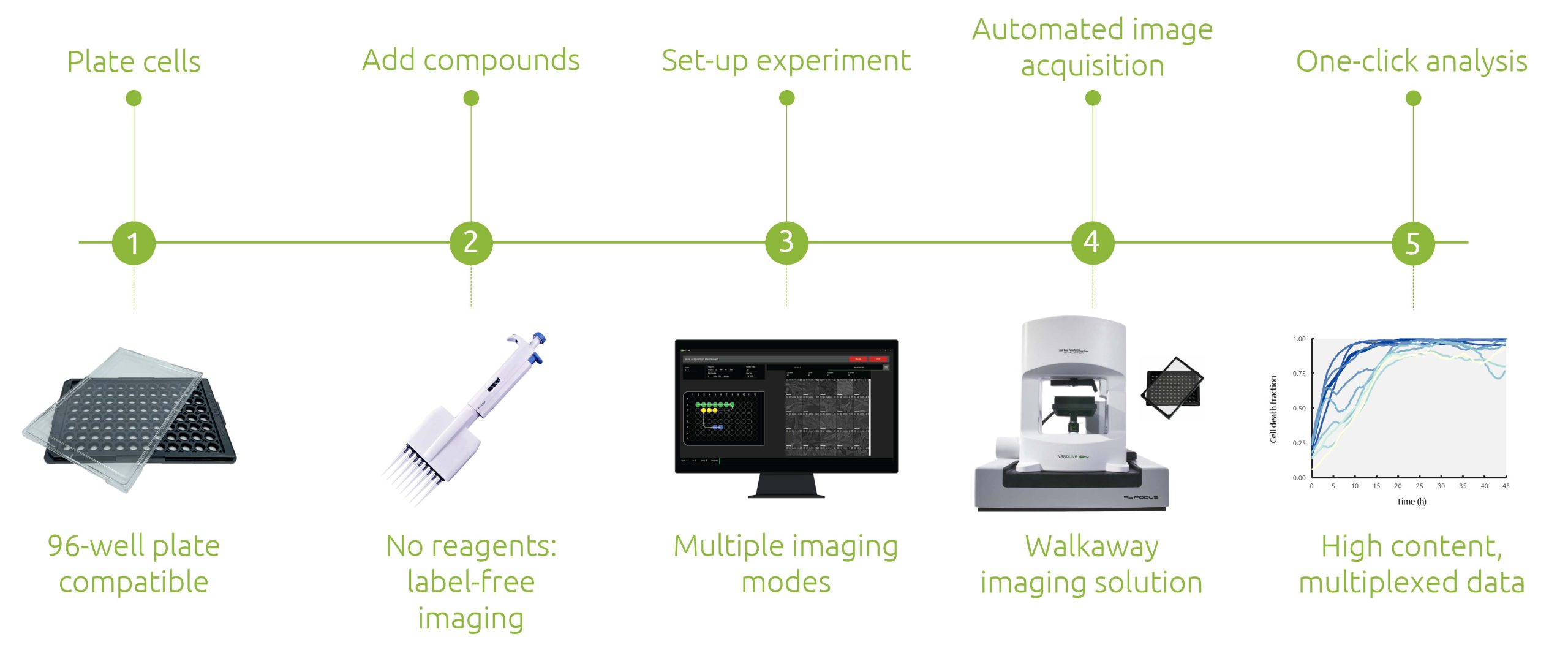
Unlock the power of unlimited high content live cell analysis with the 3D Cell Explorer 96focus. Our unique approach enables analysis and re-analysis of label-free image data with our AI-powered digital assays. As always, the simple, autonomous workflow ensures that your precious time in the lab is used efficiently, and you can be confident that your samples are in good hands, whether you are imaging for an hour, or days.
Streamlined, automated workflows: save time and money
Long-term monitoring of cells label-free
High content, multiplexed, reliable live cell data
Fully integrated, cutting-edge digital analysis solutions
Streamlined, automated workflows: save time and money
De-risk your in vitro pre-clinical studies, reduce time to failure and increase confidence in lead candidates.
Achieve efficient and cost-effective cell-based experiments
With Nanolive’s 96-well capacity and its dynamic high content unbiased data, reduce the number of separate or parallel experiments needed, thereby reducing the use of expensive cell lines and reagents
Maximize research efficiency with automated acquisition and analysis
With Nanolive’s fully automated solution combined with its multiple digital assays, use automated acquisition and analysis to reduce the hands-on time needed to run experiments. Nanolive’s data never expires!
Walk away
Nanolive’s walkaway label-free imaging solution saves hands-on time and allows you to set your acquisition and analysis running and return when imaging is completed
Long-term monitoring of cells label-free
Your live cell studies do not have to be restricted by reagents or cell properties. Nanolive’s label-free technology allows frequent image capture for days while the stage-top incubator takes care of your cells. Even fragile and sensitive cells can be imaged for long durations without fixing, damage, or phototoxicity. Capturing complex dynamic processes like neural network formation, mitochondrial dynamics, stem cell differentiation, and cell-cell killing in detail is now possible.
High content, multiplexed, reliable live cell data
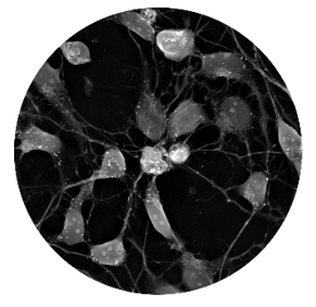
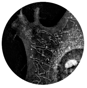
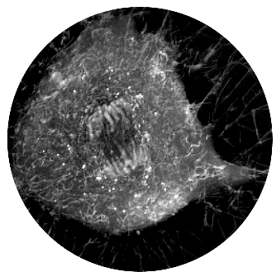
Do not miss anything! Nanolive’s technology ensures that you can switch between the population, single cell, and organelle levels when visualizing or analyzing your data without missing any detail as the sub-cellular resolution is preserved despite the field of view imaged.
Population level
At the population level, monitor cell health, cell death, apoptosis, necrosis, cell growth and cell-cell interactions
Single cell level
At the single cell level, you can analyze organelle distribution, cell morphology, and membrane dynamics
Organelle level
At the organelle level, you can observe organelle dynamics, mitochondrial morphology, chromosome condensation, and lipid accumulation
Fully integrated cutting-edge digital analytical solutions
Get higher significance earlier and faster with our panel of application specific, push-button digital assays. Retroactive analysis is also possible at any time.
Profile cell health and death, and distinguish between living, apoptotic, and necrotic cells label-free.
Measure and characterize simultaneously how live T cells find, bind, stress, and serial kill their targets.
Measure lipid droplet characteristics at population, cellular, and individual droplet levels.
Nanolive imaging and analysis platforms
Swiss high-precision imaging and analysis platforms that look instantly inside label-free living cells in 3D
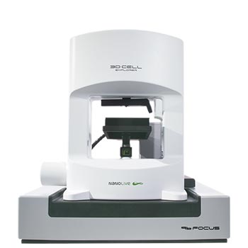
3D CELL EXPLORER 96focus
Automated label-free live cell imaging and analysis solution: a unique walk-away solution for long-term live cell imaging of single cells and cell populations
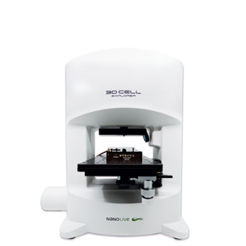
3D CELL EXPLORER-fluo
Multimodal Complete Solution: combine high quality non-invasive 4D live cell imaging with fluorescence
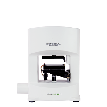
3D CELL EXPLORER
Budget-friendly, easy-to-use, compact solution for high quality non-invasive 4D live cell imaging

