In November 2020 Nanolive attended the Spanish & Portuguese Advanced Optical Microscopy (SPAOM) meeting, held remotely from Valencia in Spain. The objective of this annual congress is to keep new researchers in the Iberian Peninsula at the forefront of optical microscopy and image analysis. There is a strong focus on hands-on workshops and demonstrations at the event, which Nanolive were delighted to be a part of.
Seven researchers from across the peninsula agreed to share the data obtained with Nanolive live cell imaging, and their opinions on the insights gained with our instruments. We will be updating this blogpost every time we publish a new video.
Eduarda Martins, ICVS in Braga, Portugal
Last but definitely not least, we are very excited to promote the work of PhD student Eduarda Martins from the Life and Health Sciences Research Institute (ICVS) in Braga, Portugal. Eduarda works in Prof. Bruno Costa’s lab, which specializes in studying the molecular and cellular hallmarks of human brain tumors. Eduarda is using Nanolive imaging to observe how immune cells interact with cancer cells in real time.
In this video, we observe glioma cells interacting with immune cells in co-culture; one interesting mechanism to study tumor immune evasion. But this is not the only application that Nanolive imaging will be used for in Braga, as is explained in the video by fellow PhD student Liliana Santos. Exciting stuff! Enjoy the video for more.
Rita Soares, IMM in Lisbon, Portugal
The fourth video in this special series comes from PhD student Rita Soares, who is based at the Instituto de Medicina Molecular (IMM) in Lisbon, Portugal. Rita works in a partnership between two labs, the Mitochondria Biology & Neurodegeneration lab and the Neuronal Communication & Synaptopathies lab, under the tutelage of Dr. Vanessa Morais and Dr. Sara Xapelli. In her work, Rita uses neurosphere cultures as a means of investigating neural precursor cells in vitro. In this video, we observe a neurosphere culture from a transgenic mouse model that has been genetically-modified to express bright green fluorescent mitochondria. Mitochondria can be seen shuttling between neurons, and undergoing fission-fusion events.
To learn more about the role these processes play in cell health and the value Nanolive imaging holds for mitochondrial research, watch the video on the right.
João Martins, INL, Braga, Portugal
The third video, taken by João Martins, currently a Research Fellow at the International Iberian Nanotechnology Laboratory (INL) in Braga, Portugal, is one of our absolute favorites, and it seems to be one of yours too as the first time this video was posted on social media it went viral earning more than 400,000 views and 3500 likes. The video shows just how adept cancer cells are at evading attack from our immune cells (in this case T-cells) and is a powerful visual reminder of why cancer is such a difficult disease to beat. In the words of João Martins “cancer cells evade them (T-cells) by not being recognized, like having the invisibility cloak from Harry Potter”.
To learn more about the cell lines used in the video and the objectives of João Martins research, watch the video.
Dr. Caroline Mauvezin, IDIBELL in Barcelona, Spain
The second video comes from Dr. Caroline Mauvezin, from the Bellvitge Biomedical Research Institute (IDIBELL) in Barcelona. Dr. Mauvezin is a cell biologist and microscopy expert who specializes in autophagy and lysosome biology. She is particularly interested in understanding the roles these processes play in degenerative processes such as aging and cancer and aims to use Nanolive imaging to investigate the fate of chromosomally unstable cells which display a newly discovered atypical nuclear phenotype called the toroidal nucleus. In this video, we observe human bone osteosarcoma epithelial (U2OS) cells, genetically modified to express Histone 2B-GFP (a chromosome marker), and LAMP1-RFP (a lysosome marker).
Enjoy the explanation of her research and watch her amazing video.
Inês Faleiro, IMM in Lisbon, Portugal
The first video we will feature was obtained by PhD student Inês Faleiro, from Sérgio de Almeida’s lab at the Instituto de Medicina Molecular (IMM) in Lisbon, Portugal.
Inês uses bioimaging techniques extensively in her research and was thrilled by the results obtained with Nanolive.
Watch the video of her myotube samples and listen to her testimonial.
Read our latest news
Revolutionizing lipid droplet analysis: insights from Nanolive’s Smart Lipid Droplet Assay Application Note
Introducing the Smart Lipid Droplet Assay: A breakthrough in label-free lipid droplet analysis Discover the power of Nanolive's Smart Lipid Droplet Assay (SLDA), the first smart digital assay to provide a push-button solution for analyzing lipid droplet dynamics,...
Food additives and gut health: new research from the University of Sydney
The team of Professor Wojciech Chrzanowski in the Sydney Pharmacy School at the University of Sydney have published their findings on the toxic effect of titanium nanoparticles found in food. The paper “Impact of nano-titanium dioxide extracted from food products on...
2023 scientific publications roundup
2023 has been a record year for clients using the Nanolive system in their scientific publications. The number of peer-reviewed publications has continued to increase, and there has been a real growth in groups publishing pre-prints to give a preview of their work....
Nanolive microscopes
CX-A
Automated live cell imaging: a unique walk-away solution for long-term live cell imaging of single cells and cell populations
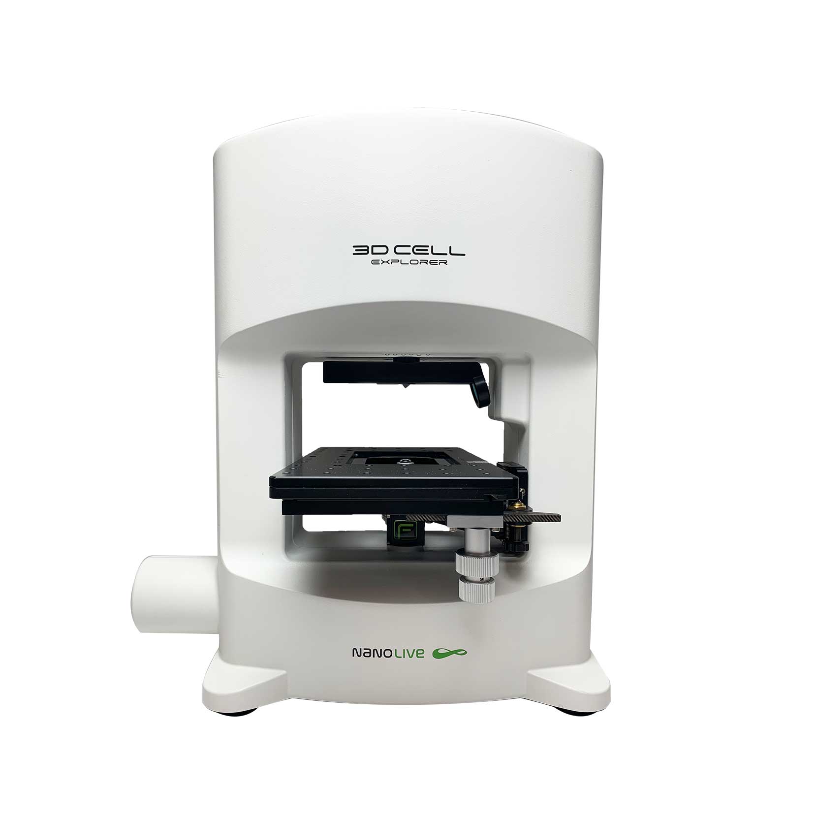
3D CELL EXPLORER-fluo
Multimodal Complete Solution: combine high quality non-invasive 4D live cell imaging with fluorescence
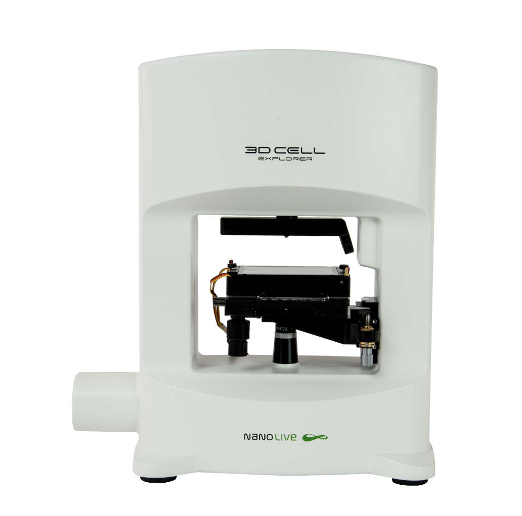
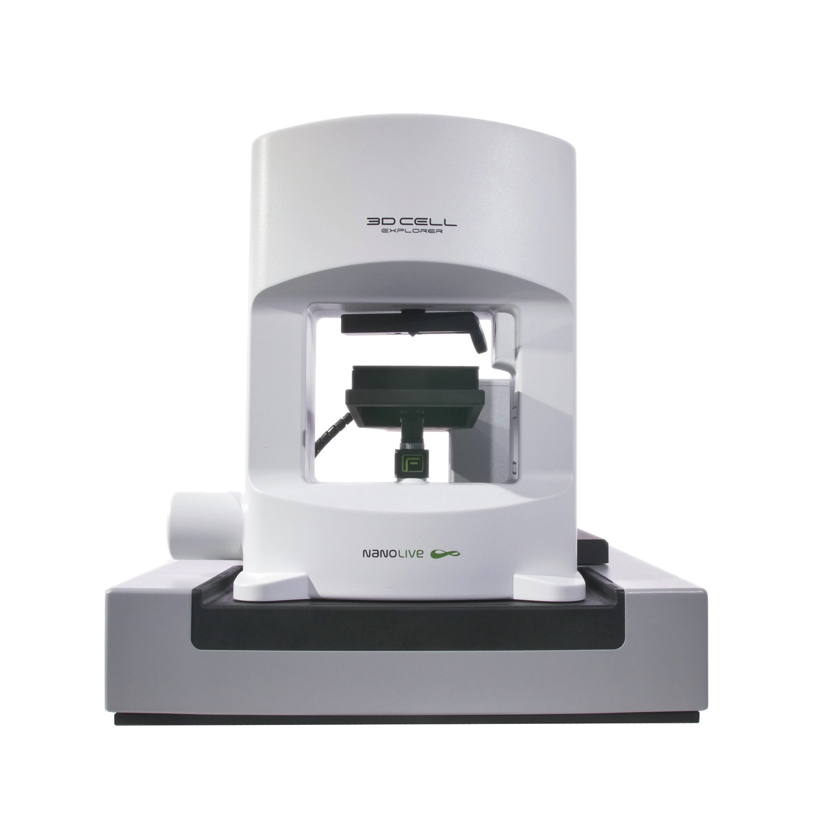
3D CELL EXPLORER
Budget-friendly, easy-to-use, compact solution for high quality non-invasive 4D live cell imaging

