Single cell characterization
Long-term label-free imaging of single cell phenotypes and cell organelles Request a demo or quoteLong-term, label-free imaging of unperturbed single cells at a population level
In this video, an unperturbed population of 3T3-derived pre-adipocyte cells were imaged for 90 h using the 10×10 grid-scan mode on Nanolive’s 3D Cell Explorer 96focus (one image taken every 8.5 mins).
Watch as confluence increases from 9% to 60% over the course of the video (from 74 cells to 521) and marvel at the precision of the segmentation masks that make calculations like this possible.
Quantifying phenotypic variability at the population level
Phenotypic variation is a ubiquitous feature of all biological systems. It underpins complex, dynamic systems (e.g. immune responses) and biological processes (e.g. stem cell differentiation). Measuring changes in heterogeneity requires multiplex imaging platforms that maintain high spatial and temporal resolution, like Nanolive’s 3D Cell Explorer 96focus. Here, we provide the first quantitative analysis of phenotypic variability of single cells in a population of unperturbed cells.
Here, three cell metrics (cell area, dry mass, and compactness) are calculated for each cell, at every time point and visualized as relative frequency histograms. This approach allows to measure micro-heterogeneity in a population and to compare the variance that exists between traits.
Multiplex organellular imaging
Nanolive’s high resolution allows the behaviour and dynamics of multiple cell organelles to be measured at the same time, without the addition of labels. In this video, we show close-up footage of the dynamics of five organelles (nucleus, mitochondria, lipid droplets, lysosomes, vesicles) and membrane protrusions in primary human keratinocytes. Discover here our cell organelles page.
Download here our poster “Quantifying dry mass dynamics in lipid droplets using Nanolive cell imaging”.
Capturing and quantifying novel biological phenomena such as nuclear spinning
Researchers from EPFL have used Nanolive cell imaging to capture, and quantify, the dynamic behaviour of cellular organelles. Their findings, published in leading journal PLOS Biology, include an in-depth, quantitative analysis of the dry mass dynamics of mammalian organelles, and nuclear spinning. Download here the PLOS Biology paper.
Scientific Publications
Nanolive label-free live cell imaging has already shed light on many important topics in the field of single cell characterization research. To get inspired and learn how your research can benefit from our technology, we invite you to check out these scientific articles published by our clients.
Feature Application

Feature application: Characterization of single cells
In this Feature Application, we showcase the enormous potential that Nanolive live cell imaging holds for single cell characterization. In the first case study, we analyze micro-heterogeneity at the population and the temporal level in unperturbed cells and in the second and third case studies we focus on cells where intracellular trafficking and respiration were experimentally perturbed.
Webinar 1
Watch our webinar: Label-free analysis of living cell populations reveals controlled phenotypic variation among single cells
In this webinar, Dr. Emma Gibbin-Lameira, Communications Specialist at Nanolive:
- Compares the variability of isolated single cells with the variability of single cells in a large population to determine whether heterogeneity is driven by internal or external factors.
- Examines how the variability of individual traits change over time in unperturbed cells.
- Analyzes how cells respond to perturbations in intracellular trafficking and respiration to determine whether targeting different systems induces/reduces the phenotypic variation of single cells in a population.
Please register here to view the webinar on-demand:
Webinar 2
Watch our webinar: Label-free quantification of cell metabolism – focus on mitochondria and lipid droplets dynamics
In this webinar, Dr. Emma Gibbin-Lameira, Communications Specialist at Nanolive will:
- Show time-lapse footage of mitochondrial swelling and dysfunction following perturbation.
- Introduce a cell metric called granularity and discuss whether changes in this measurement could be a signal of stress preceding cell death.
- Present a fully quantitative analysis of lipid droplet dynamics in unperturbed cells, starting at the population level and finishing at level of an individual lipid droplet. Correlations between lipid droplet characteristics and other cell morphological traits (e.g. dry mass) will also be featured for the first time.
Please register here to view the webinar on-demand:
Video Library
Nanolive imaging and analysis platforms
Swiss high-precision imaging and analysis platforms that look instantly inside label-free living cells in 3D

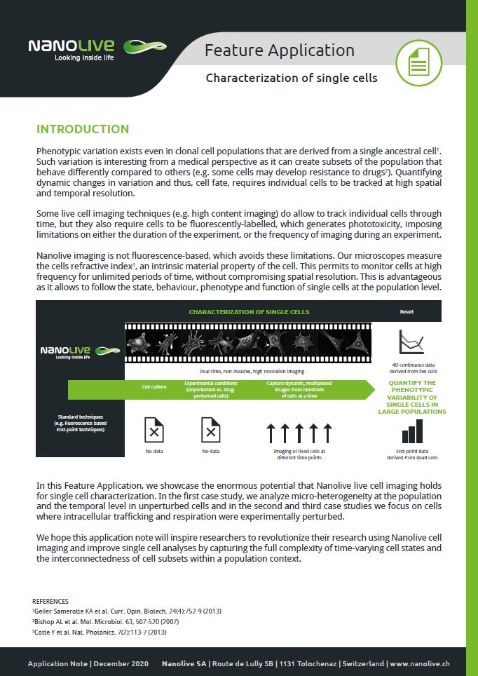
3D CELL EXPLORER 96focus
Automated label-free live cell imaging and analysis solution: a unique walk-away solution for long-term live cell imaging of single cells and cell populations

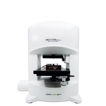
3D CELL EXPLORER-fluo
Multimodal Complete Solution: combine high quality non-invasive 4D live cell imaging with fluorescence

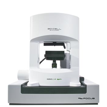
3D CELL EXPLORER
Budget-friendly, easy-to-use, compact solution for high quality non-invasive 4D live cell imaging
