Microbiology & host-pathogen interactions
Long term non-invasive live cell imaging of bacteria, viruses, fungi, and protozoa Request a demo or quoteInvestigating COVID-19 infection
This video (images acquired every 10 mins for 20 h), shows the shocking impact that SARS-CoV-2 has on cell fate. Infected cells fuse with neighbouring cells, forming large multi-nucleated cells; a process known as syncytia formation. This footage, captured by post-doctoral researcher Dr. Blandine Monel, was kindly provided by Professor Olivier Schwartz from the Virus and Immunity lab at the Pasteur Institute in Paris.
Read the blogpost here.
Testing a compound’s antimicrobial activity
In this video, we observe the response of Streptococcus pneumoniae to the endolysin, ClyJ-3. The disappearance of the bright spots in the refractive index images indicates that, as expected, most bacteria are killed by ClyJ-3. One image was taken every min for 2 h using the 3D Cell Explorer. This video was kindly provided by Prof. Hak-Kim Chan from the Faculty of Medicine and health at the University of Sydney (Australia).
Read the blogpost here.
Capturing virus-induced cytopathic effects
In this footage, obtained using Nanolive’s 3D Cell Explorer-fluo we observe the progression of GFP-labelled adenovirus in human bone osteosarcoma epithelial (U2OS) cells. Refractive images were taken every 15 s and fluorescence images were taken every 5 min using the FITC channel, for a total of 25 mins. This video was kindly provided by Prof. Clodagh O’Shea from the Salk Institute for Biological Studies (La Jolla, CA, USA). Read our interview with Prof. O’Shea here.
Visualizing fungal spore germination
In this footage, which was obtained during a demonstration organized by Dr. Matthias Steiger at the Technical University in Vienna, we observe the germination process in Trichoderma reesei at 30 °C. One image was acquired every 5 min for 2 h 45 min using Nanolive’s 3D Cell Explorer. This time frame was sufficient to observe the initial formation of the germ tube. The high resolution of the images permit visualization of the growing membrane, internal structures and the formation of the wall, which occurs after 2 h 20 min.
Observing the progression of the intra-erythrocytic cell cycle of protozoans
To infect its host, the parasite Plasmodium falciparum must avoid macrophage detection, and degrade huge amounts of haemoglobin. This loss of haemoglobin is visible under the 3D Cell Explorer.
In this video, we observe the morphological changes induced by P. falciparum infection at different stages of the parasite’s life cycle.
Read more in our detailed blogpost here.
Scientific publications
Video library
Nanolive imaging and analysis platforms
Swiss high-precision imaging and analysis platforms that look instantly inside label-free living cells in 3D

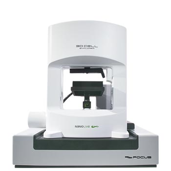
3D CELL EXPLORER 96focus
Automated label-free live cell imaging and analysis solution: a unique walk-away solution for long-term live cell imaging of single cells and cell populations

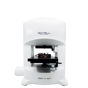
3D CELL EXPLORER-fluo
Multimodal Complete Solution: combine high quality non-invasive 4D live cell imaging with fluorescence

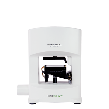
3D CELL EXPLORER
Budget-friendly, easy-to-use, compact solution for high quality non-invasive 4D live cell imaging
