Professor Olivier Schwartz heads a 14-person strong laboratory at the Virus and Immunity lab at the prestigious Pasteur Institute in Paris. His lab studies how pathogenic viruses spread and interact with the host immune system. Before 2020, Prof. Schwartz’ research focused primarily on HIV, Zika, and dengue viruses, but the COVID-19 pandemic forced him to change track and focus on the SARS-CoV-2 infection process. His team are interested in understanding how the virus enters cells and what happens once a cell becomes infected. Nanolive’s unique capacity to capture the dynamic responses of living cells to viruses convinced Prof. Schwarz to book a demonstration, and so a CX-A was installed in their P3 L3 facility.
Prof. Schwartz and post-doctoral researcher, Dr. Blandine Monel set up an experiment designed to test whether Nanolive technology could capture the response of cells to SARS-CoV-2 infection and “were very lucky to observe striking changes mediated by the virus”. The team were convinced and bought a CX-A several months ago. We asked Prof. Schwartz whether he was willing to share their preliminary results with us, and he kindly agreed. Watch the full interview in the video below.
The video on the right (images acquired every 10 mins for 20 h), shows the shocking impact that SARS-CoV-2 has on cell fate. Infected cells fuse with neighbouring cells, forming large multi-nucleated cells; a process known as syncytia formation. This response is not specific to viruses belonging to the Coronaviridae; several viral families induce this form of cell-cell fusion, including the Herpesviridae (responsible for among others HIV) and the Paramyxoviridae (best known as the cause of measles)1. Syncytia express higher amounts of viral antigens in the tissues and organs of infected hosts2. As SARS-CoV-2 viral load is associated with increased disease severity and mortality3, there is great interest in investigating the role that syncytia play in COVID-194.
Prof. Schwartz’ lab plan on using the “unprecedented details that Nanolive imaging provide about the cell cellular organization, the organelles, the nuclei, the nucleoli, the cytoskeleton, the mitochondria, and the lipid droplets” to investigate the consequences cell-cell fusion has for the overall organization of the cell, particularly on the mitochondrial behaviour within the cells. They also plan on studying the host immune response to the infection; how antibodies counteract viral replication and neutralize cell-cell fusion.
Nanolive would like to thank Prof. Schwartz for letting us share these exciting preliminary results. His team is currently busy reproducing the experiments to validate the results, but this is certainly an interesting avenue for research.
References
1Leroy H. et al. 2020. Int. J. Mol. Sci. 21(24):9644.
2Hijano DR et al. 2019. PloS One. 3;14(9):e0220908.
3Fajnzylber J. et al. 2020. Nat. Commun. 30;11(1):1-9.
4Buchrieser J. et al. 2020. EMBO J. 1;39(23):e106267.
Read our latest news
Revolutionizing lipid droplet analysis: insights from Nanolive’s Smart Lipid Droplet Assay Application Note
Introducing the Smart Lipid Droplet Assay: A breakthrough in label-free lipid droplet analysis Discover the power of Nanolive's Smart Lipid Droplet Assay (SLDA), the first smart digital assay to provide a push-button solution for analyzing lipid droplet dynamics,...
Food additives and gut health: new research from the University of Sydney
The team of Professor Wojciech Chrzanowski in the Sydney Pharmacy School at the University of Sydney have published their findings on the toxic effect of titanium nanoparticles found in food. The paper “Impact of nano-titanium dioxide extracted from food products on...
2023 scientific publications roundup
2023 has been a record year for clients using the Nanolive system in their scientific publications. The number of peer-reviewed publications has continued to increase, and there has been a real growth in groups publishing pre-prints to give a preview of their work....
Nanolive microscopes
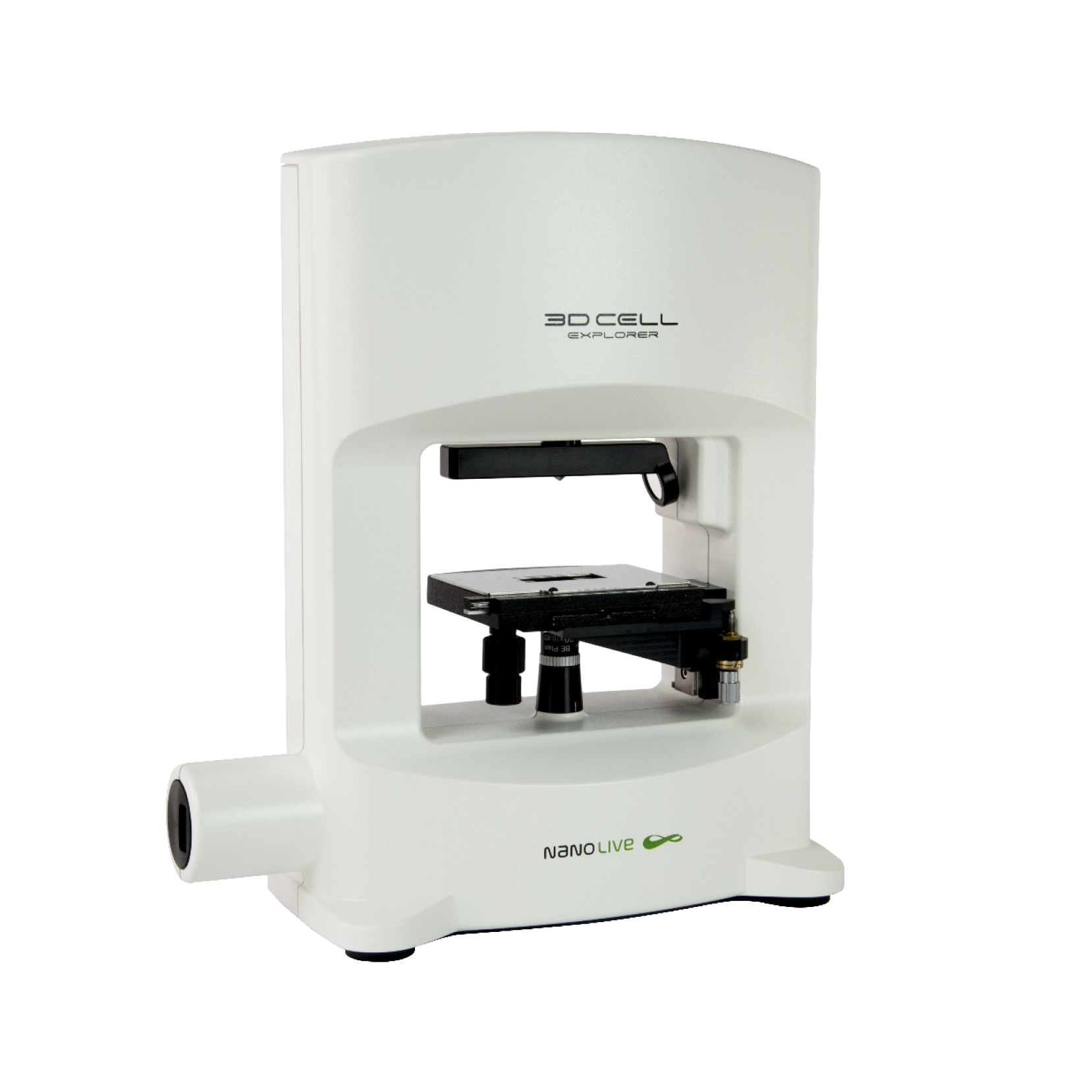
3D CELL EXPLORER
Budget-friendly, easy-to-use, compact solution for high quality non-invasive 4D live cell imaging
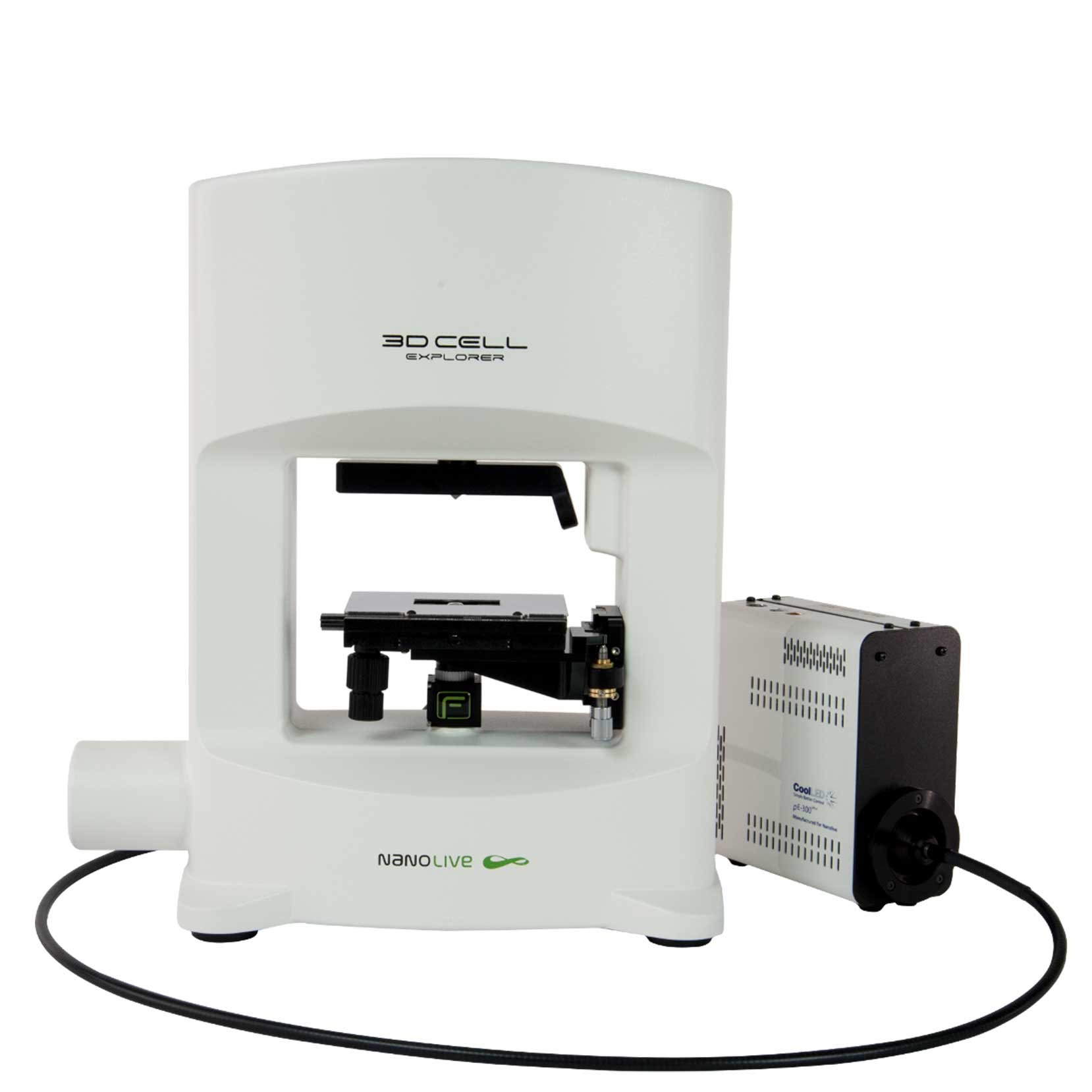
3D CELL EXPLORER-fluo
Multimodal Complete Solution: combine high quality non-invasive 4D live cell imaging with fluorescence
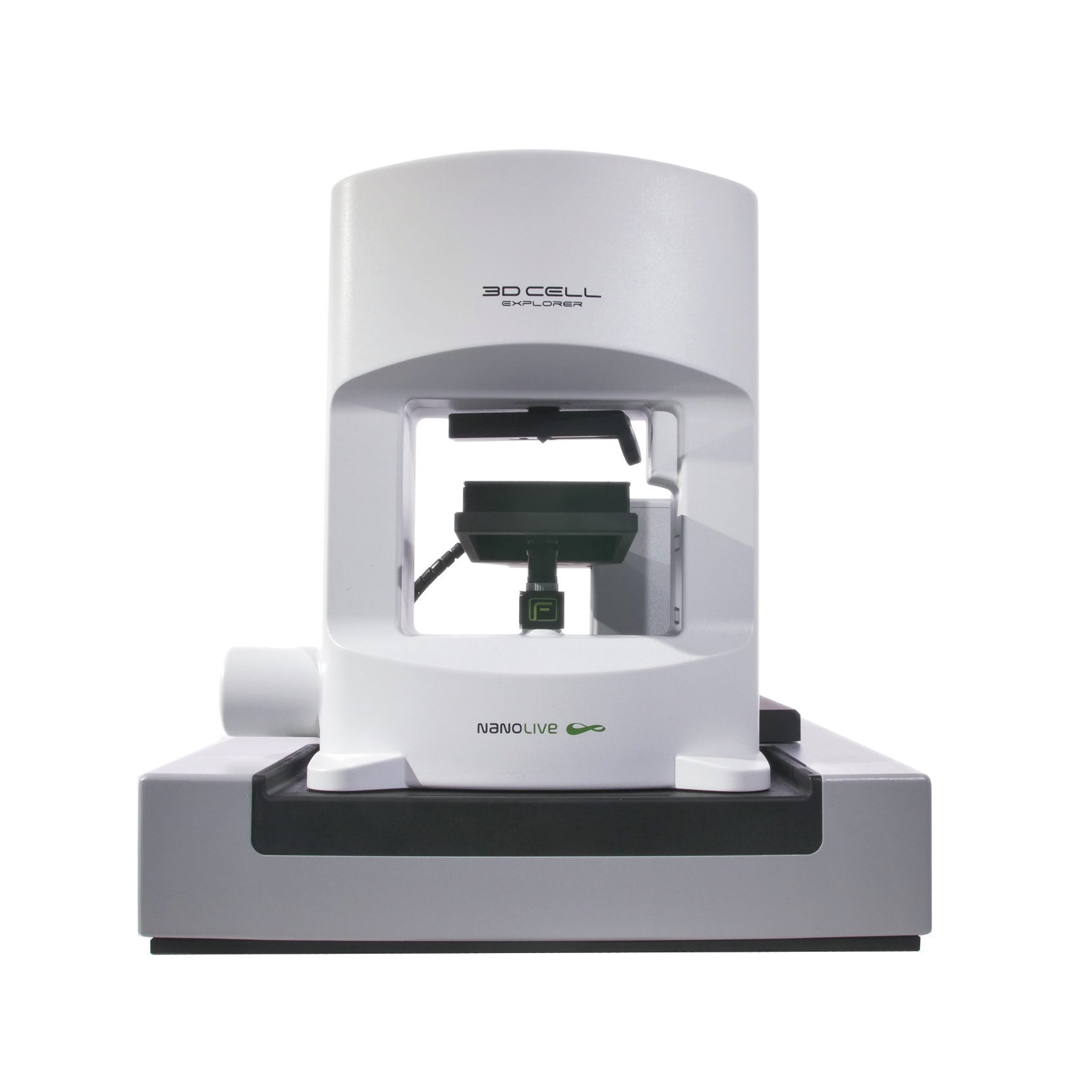
CX-A
Automated live cell imaging: a unique walk-away solution for long-term live cell imaging of single cells and cell populations

