Cytotoxicity research
Request a quote or demoScientific Publications
Nanolive label-free live cell imaging has already shed light on many important topics in the field of drug discovery research. To get inspired and learn how your research can benefit from our technology, we invite you to check out these scientific articles published by our clients.
Automating phenotypic screening
Small-molecule kinase inhibitors have had success in the treatment of many diverse types of cancer. Here, we use Nanolive’s CX-A to test how cells respond to various inhibitors or modulators of kinase activities (Akt, JNK, CDK2, RTK, PKA, JAK, mTOR and AMPK). Images were taken using the automated 3×3 gridscan mode, at an acquisition rate of 1 scan every 25 mins, for 40 h.
Read our detailed blogpost here.
Quantifying non-homogeneous population responses to drug-perturbation
Ebastine is an antihistamine that simultaneously disrupts lysosomes and mitochondria. The cell stress response to Ebastine is highly non-homogeneous; a sub-population of cells die within hours of exposure to the drug, predominantly by apoptosis, but others are still alive 20 h later. This raises an important question: what makes certain cells more resistant than others?
The sub-cellular resolution delivered by Nanolive imaging reveals more resistant cells contain small black endocytic vacuoles in their cytoplasm. This, combined with global changes in granularity; a texture measurement that describes how a signal is distributed at a certain scale, suggests there is a reorganization of the subcellular distribution of organelles during the experiment, which could explain why certain cells survive longer than others.
Watch our dedicated cytotoxicity webinar here.
Increasing biological relevance and simplifying the discovery workflow
These are the results of a case study that was created to demonstrate how the CX-A and Eve Analytics can be used in drug screening. We feature the entire process from image acquisition to data analysis and interpretation in our webinar here. In the case study, we test to what extent the art of observing influences the outcome of our experiments; something too often ignored when running live cell experiments. The video, shows the effects that four different treatments: (1) label-free; (2) cytotoxic (MitoTracker only); (3) cytotoxic and low phototoxic (MitoTracker and 1% laser power) and (4) cytotoxic and high phototoxic (MitoTracker and 5% laser power), have on cell health.
Gaining insights into a drugs mechanism of action
Taxol is an anti-cancer chemotherapy drug that disrupts the formation of mitotic spindles during metaphase. The sub-cellular resolution of the CX-A allows us to visualize this process in spectacular detail; when cells enter mitosis, they successfully complete prophase and prometaphase but are unable to line their chromosomes up along the metaphasic plate leading to failure of cytokinesis and the development of multiple nuclei per cell.
With EVE Analytics, Nanolive’s quantitative software solution, we are able to quantify the multinucleation event from the increase in average dry mass density per cell.
Watch our dedicated cytotoxicity webinar here.
Application Note
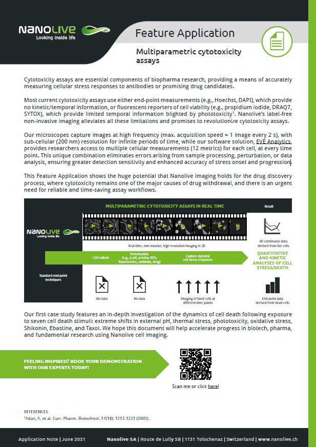
Feature Application: Multiparametric cytotoxicity assays
In this application note we demonstrate how the huge potential that Nanolive imaging holds for the drug discovery process, where cytotoxicity remains one of the major causes of drug withdrawal, and there is an urgent need for reliable and time-saving assay workflows. We show it is possible to quantify the onset and progression of stress with high precision, bringing greater accuracy to cell death research.
Webinar 1
Watch our Webinar: Do different cell death stimuli show unique signatures in cell metrics?
In this webinar, Dr. Emma Gibbin-Lameira, Communications Specialist at Nanolive reveals how Nanolive imaging can be used to conduct real-time, automated cytotoxicity assays, without the contribution of photo and cytotoxic methodological artefacts. Specifically, she will:
- Show high resolution, time-resolved footage of the dynamics of cell death following exposure to seven cell death stimuli (extreme shifts in external pH, thermal stress, phototoxicity, oxidative stress, Shikonin, Ebastine and Taxol)
- Feature relevant cell metrics and/or close-ups of changes in sub-cellular morphology induced by each experimental condition
- Discuss which cell metrics are best for distinguishing between different types of cell death
Sign up to watch the webinar:
Webinar 2
Watch our Webinar: Accelerating oncology drug discovery with label-free imaging
In this webinar, Dr. Emma Gibbin-Lameira, Communications Specialist at Nanolive will discuss the advantages of using label-free live cell imaging in the drug discovery process and show how Nanolive’s automated live cell imaging solution the CX-A, can be used to:
- observe unique drug-induced phenotypes that fluorescence microscopy cannot capture
- record dynamic cellular responses to drug perturbations
- automate in vitro screening studies
Dr. Gibbin-Lameira will present novel results from two experimental screens designed to test the phenotypic and morphological responses of pre-adipocyte and cancer cells to 8 inhibitors or modulators of kinase activity.
Sign up to watch the webinar:
Webinar Tea Break
Watch our Webinar: Tea break: Quantifying cell cytotoxicity in 96 WP format label-free
Summary: Dr. Gibbin-Lameira presents a powerful cytotoxicity case study showing Nanolive’s unique live cell-based testing and high content analysis in 96-well plate.
- Multiplexed quantitative descriptors of cell morphology and composition at both the population (field of view) and single cell level can be combined with high spatio-temporal images to evaluate drug responses in parallel, over long periods of time
- It is possible to quantify dose-response effects of drugs on living cells in a multi-well set-up where different concentrations and different drugs can be tested in parallel.
Sign up to watch the webinar:
Video Library
Nuclear changes following transcription inhibition
Read the blogpost here
Norepinephrine effect on 3T3-derived pre-adipocyte cells
Read the blogpost here
The phenotypic effects of Valproic Acid
Read the blogpost here
Cell death following exposure to oxidative stress
Watch our webinar here
Morphological noise after Rotenone-perturbation
Read our Application note here
Changes in granularity after Antimycin A addition
Read our Application note here
Single cell changes in granularity after Antimycin A addition
Read our Application note here
Staurosporine-induced cell death in HeLa cells
Erastin-induced cell death in HeLa cells
Chloroquine-induced apoptosis in HeLa cells
Cytotoxicity-induced apoptosis
Watch the webinar here
Cyto- and phototoxicity-induced necroptosis
Watch the webinar here
Cell death following extreme shifts in external pH
Watch our dedicated cytotoxicity webinar here
Cell death following extreme shifts in thermal stress
Watch our dedicated cytotoxicity webinar here
Cell death following exposure to FITC
Watch our dedicated cytotoxicity webinar here
Cell death following Shikonin addition
Watch our dedicated cytotoxicity webinar here
Quantitative analysis of the phenotypic response of HeLa cells to Staurosporine
Read our blogpost here
Compare Nanolive’s Products

CX-A
Automated live cell imaging: a unique walk-away solution for long-term live cell imaging of single cells and cell populations

3D CELL EXPLORER-fluo
Multimodal Complete Solution: combine high quality non-invasive 4D live cell imaging with fluorescence

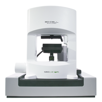
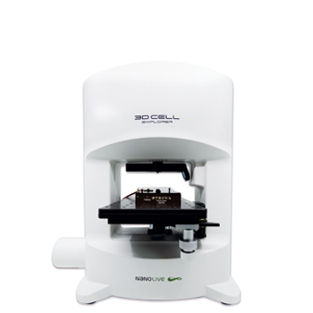
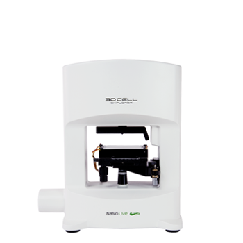
3D CELL EXPLORER
Budget-friendly, easy-to-use, compact solution for high quality non-invasive 4D live cell imaging

