Introduction to holotomography with Nanolive’s eBook
Download the eBookExploring the frontiers of label-free imaging: Nanolive’s holotomography and AI analysis
Revealing the complexities of live cell imaging for research and drug development
This eBook serves as a comprehensive guide and expansion on the innovative miniseries “Discover label-free imaging technology with Nanolive“, which introduces the fascinating world of holotomography and its groundbreaking implications for live-cell imaging.

Included inside
What is holotomography?
Nanolive imaging platforms use holotomography to capture label-free timelapse images of cells and their organelles to give insight into dynamic processes and phenotypic changes, but how does the technology work? Chapter 1 explains the concept of holotomography, and Nanolive’s unique patented approach.

Figure 1. Nanolive’s holotomography: an imaging technique combining holography and tomography.
Comparing label-free imaging methods
What’s the difference between holotomography, phase contrast, and quantitative phase imaging (QPI)? These three imaging techniques are all based on the same label-free principle but can provide differing levels of resolution. Chapter 2 compares these technologies and their uses in live-cell imaging, from cell counting, to high-content phenotypic analysis.

Figure 2. Side-by-side image comparison between different label-free imaging technologies. Phase contrast imaging enables label-free cell counting and basic morphological analysis at the cellular level. Quantitative phase imaging reduces the halo effect seen with phase contrast, and allows analysis of cellular dry mass, and more morphological details. Nanolive’s holotomography achieves high resolution, high-contrast images in 3D, allowing in-depth analysis from the population, to the organelle level, and even enabling detection of specific processes like apoptosis, or T cell-target cell interaction, without labels.
Nanolive’s unique approach to holotomography
Nanolive’s imaging platforms were developed from the ground up, incorporating unique patented components. Chapter 3 highlights the key features that set Nanolive’s holotomographic imaging apart in our mission to give researchers access to unique live cell data through AI-assisted analysis, to accelerate and de-risk the discovery and development of therapeutics.

Figure 3. Cytotoxicity screen in a 96-well plate, using Nanolive’s LIVE Cytotoxicity Assay.
Label-free high-content imaging with Nanolive’s AI-powered analysis
The high resolution of Nanolive images enables a deep understanding of cellular processes. With the help of automated AI-powered assays, phenotypic changes can be measured and extracted to quantify cellular response to drugs, cell-cell interactions, and cell death to name just a few. Chapter 4 demonstrates how Nanolive’s digital assays can be used to quantify phenotypic changes from label-free images, providing insights for fundamental research and drug development.

Figure 4. Overview of Nanolive’s suite of automated digital assays for cell analysis.
Watch the accompanying video miniseries
Image references
All used under Creative Commons license CC BY 4.0 https://creativecommons.org/licenses/by/4.0/
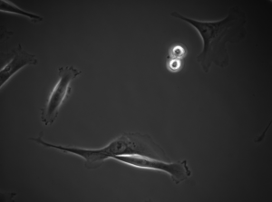 |
Ayanzadeh, A., Yağar, H.O., Özuysal, Ö.Y., Okvur, D.P., et al. (2019) Phase Contrast Microscopy of cells with annotation. Frame 470 (Cropped from original). Available from: https://doi.org/10.6084/m9.figshare.8965820.v2 |
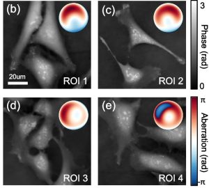 |
Shu, Y., Sun, J., Lyu, J. et al. Adaptive optical quantitative phase imaging based on annular illumination Fourier ptychographic microscopy. PhotoniX 3, 24 (2022). (Cropped from original). Available from: https://doi.org/10.1186/s43074-022-00071-3 |