Mesenchymal stem cells (MSCs) hold great promise for regenerative medicines such as cell therapy and tissue engineering (1,2). These approaches require cells to be grown in culture and differentiated into specific cell types such as osteoblasts (bone cells), chondrocytes (cartilage cells), myocytes (muscle cells), adipocytes (fat cells) or neurons (nerve cells). Here, we use Nanolive’s automated microscope, the CX-A, to make novel observations about the various stages involved in neuronal differentiation.
Human MSCs were grown in low-serum cell growth media in 35 mm dishes pre-coated with fibronectin. The first video in the compilation shows a healthy population of MSCs undergoing mitosis (00:00 to 00:17). At this pre-differentiation stage cells are motile, and their (large, flat) morphology is homogenous.
When neurogenic cell differentiation media was added the cells stop dividing (00:17 to 00:42). No acute changes in morphology were observed over the first couple of hours, but after approximately 6 h differentiation starts to occur. Most cells have the long, thin, spindle-shaped morphology of neural progenitor cells, while a small minority assume the radial morphology of glial cells (00:42 to 00:59).
Differentiation success appears to be related to confluency: at low confluency, cells are highly mobile and seem to differentiate less. Conversely, at high confluency almost all cells show morphological signs of differentiation (00:59 to 01:28).
Several days after the differentiation media is added, more complex, advanced changes in morphology become visible (01:28 to 02:18). These changes in morphology are a consequence of the retraction of the actin cytoskeleton. Branched protoplasmic extensions resembling dendrites are formed, some of which (specifically those connected to neighboring cells) eventually form axons.
References
(1) Xu, Y., Shi, Y. and Ding, S. 2008. A chemical approach to stem-cell biology and regenerative medicine. Nature, 453(7193), 338-344.
(2) Wu, S.M. and Hochedlinger, K. 2011. Harnessing the potential of induced pluripotent stem cells for regenerative medicine. Nature Cell Biology, 13(5), 497-505.
Read our latest news
Cytotoxic Drug Development Application Note
Discover how Nanolive’s LIVE Cytotoxicity Assay transforms cytotoxic drug development through high-resolution, label-free quantification of cell health and death. Our application note explores how this advanced technology enables real-time monitoring of cell death...
Investigative Toxicology Application Note
Our groundbreaking approach offers a label-free, high-content imaging solution that transforms the way cellular health, death, and phenotypic responses are monitored and quantified. Unlike traditional cytotoxicity assays, Nanolive’s technology bypasses the limitations...
Phenotypic Cell Health and Stress Application Note
Discover the advanced capabilities of Nanolive’s LIVE Cytotoxicity Assay in an application note. This document presents a detailed exploration of how our innovative, label-free technology enables researchers to monitor phenotypic changes and detect cell stress...
Nanolive microscopes

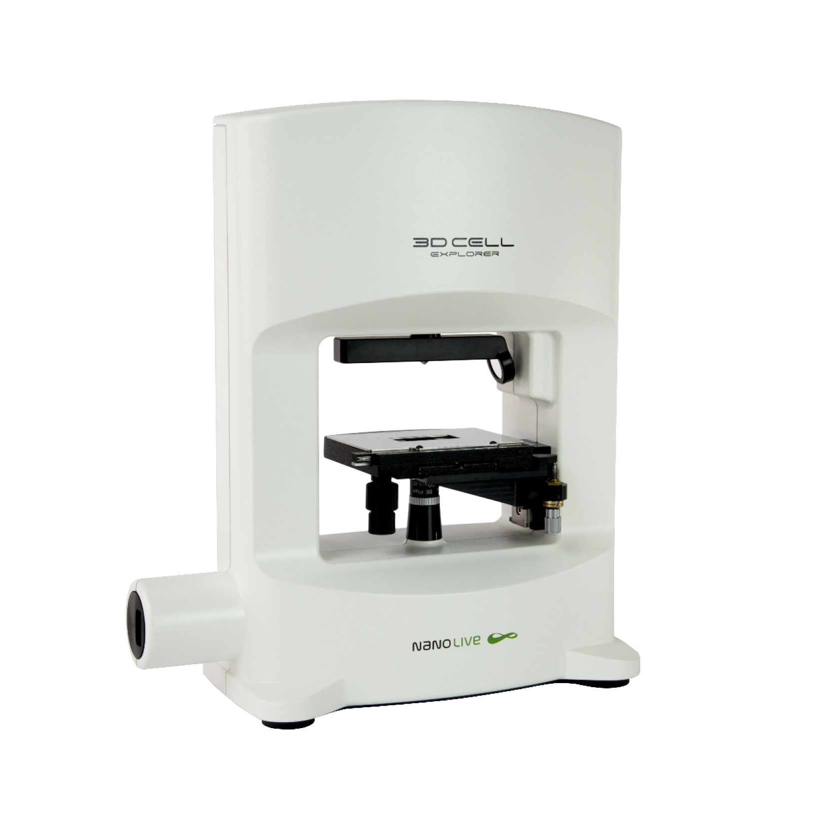
3D CELL EXPLORER
Budget-friendly, easy-to-use, compact solution for high quality non-invasive 4D live cell imaging

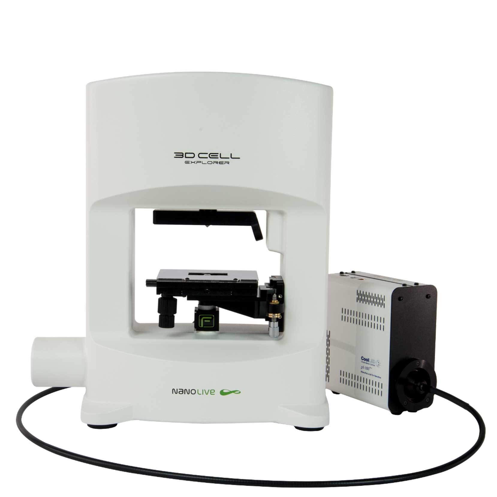
3D CELL EXPLORER-fluo
Multimodal Complete Solution: combine high quality non-invasive 4D live cell imaging with fluorescence

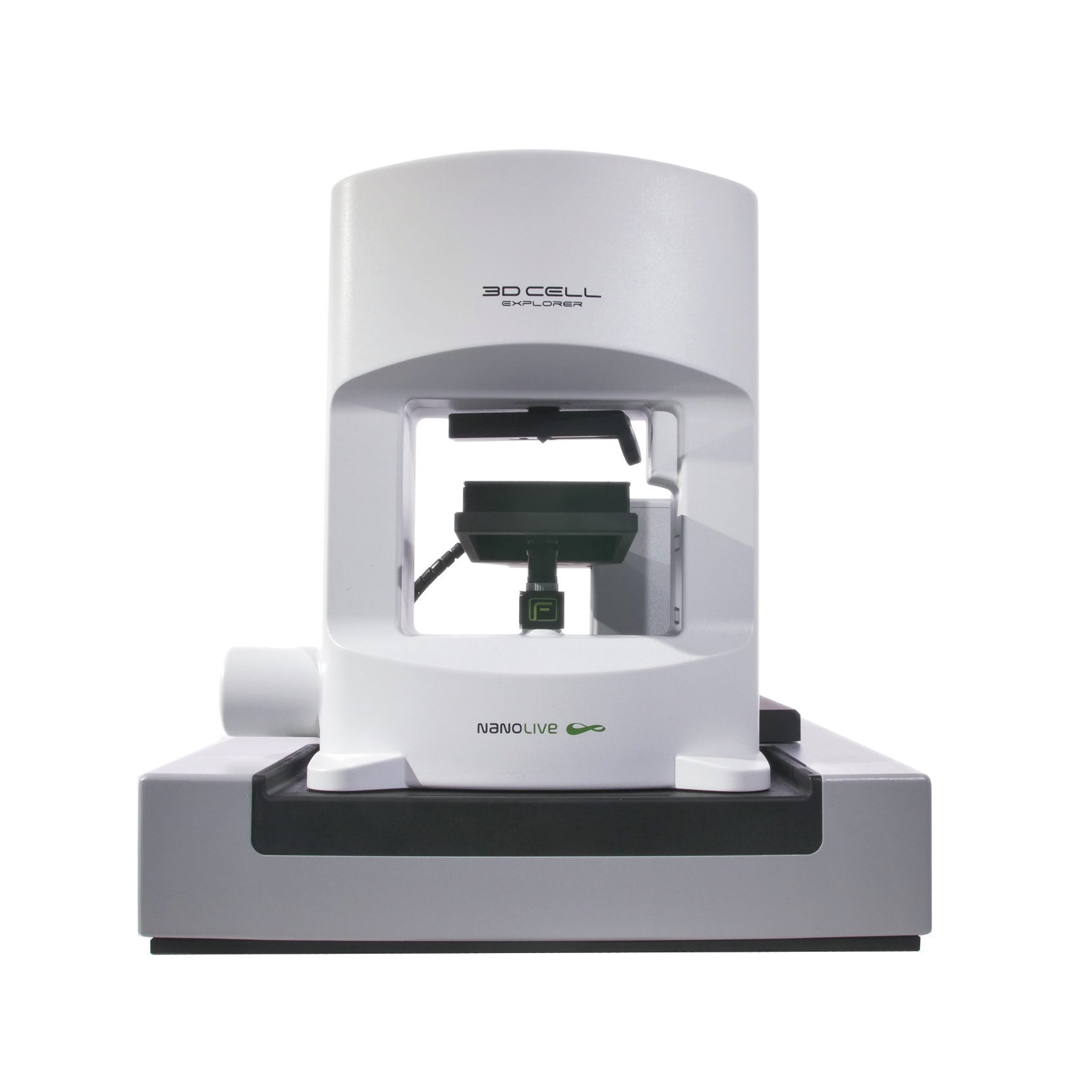
CX-A
Automated live cell imaging: a unique walk-away solution for long-term live cell imaging of single cells and cell populations


