We are happy to announce a new publication in the Journal of Physics from users of the Nanolive’s 3D Cell Explorer in the Ioffe Institute in St. Petersburg, Russia.
Photodynamic therapy is a common treatment against some types of cancer, like esophageal cancer and non-small cell lung cancer. It consists on the administration of a drug that, once exposed to a specific wavelength of light, produces reactive oxygen species that lead to the death of nearby cells.
The investigation of living cells response to photodynamic treatment is crucial to understand drug pharmacodynamics and to widen the targeted cancer types.
Gorbenko, D. A and colleagues investigated post-treatment dynamics in living cells under the 3D Cell Explorer[1].
HeLa cells were treated with a photosensitizer drug at different concentrations. After treatment, the cells were irradiated at different doses with the specific wavelength for the drug.
Three conditions were studied: low treatment dose (low drug concentration and low irradiation dose), moderate dose (high drug concentration and moderate irradiation dose), and high dose (high drug concentration and high irradiation dose).
The formation of reactive oxygen species as a result of the irradiation of the drug produces changes in cell volume, cell average height and cell membrane area. These changes were observed under the 3D Cell Explorer and quantified.
The results showed that, at the low dose, cells resisted the photodynamic treatment, and no changes could be observed 24h after treatment.
At the moderate dose, the experimenters could obverse blebbing, cell rounding, a noticeable decrease in cell volume and a decrease in membrane area. Those cells underwent apoptosis.
Finally, at the higher dose, cells showed membrane rupture and leakage of the intracellular content, which are typical signs of necrosis.
The results were validated with confocal fluorescence microscopy.
The 3D Cell Explorer again proved to be a tool of choice in cytotoxity and cellular compound studies. Their full publication is available here!
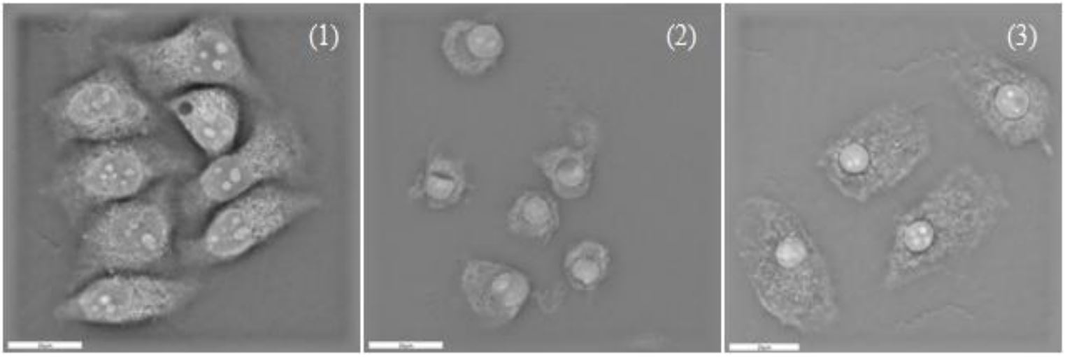
Figure 1. Holographic tomography Z-cross sections of HeLa cells subjected to PD treatment at: (1) Irradiation dose 5 J, PS concentration 5 µg/ml; (2) 10 J, 10 µg/ml; (3) 20 J, 10 μg/ml. Image from D A Gorbenko et al 2019 J. Phys.: Conf. Ser. 1236 012015.
[1] D. A. Gorbenko, A. V Belashov, T. N. Belyaeva, E. S. Kornilova, I. V Semenova, and O. S. Vasyutinskii, “Quantification of changes in cellular morphology during cell necrosis obtained from 3D refractive index distributions,” p. 12015, 2019.
Read our latest news

Revolutionizing lipid droplet analysis: insights from Nanolive’s Smart Lipid Droplet Assay Application Note
Introducing the Smart Lipid Droplet Assay: A breakthrough in label-free lipid droplet analysis Discover the power of Nanolive's Smart Lipid Droplet Assay (SLDA), the first smart digital assay to provide a push-button solution for analyzing lipid droplet dynamics,...

Food additives and gut health: new research from the University of Sydney
The team of Professor Wojciech Chrzanowski in the Sydney Pharmacy School at the University of Sydney have published their findings on the toxic effect of titanium nanoparticles found in food. The paper “Impact of nano-titanium dioxide extracted from food products on...

2023 scientific publications roundup
2023 has been a record year for clients using the Nanolive system in their scientific publications. The number of peer-reviewed publications has continued to increase, and there has been a real growth in groups publishing pre-prints to give a preview of their work....
Nanolive microscopes

3D CELL EXPLORER
Budget-friendly, easy-to-use, compact solution for high quality non-invasive 4D live cell imaging

3D CELL EXPLORER-fluo
Multimodal Complete Solution: combine high quality non-invasive 4D live cell imaging with fluorescence

CX-A
Automated live cell imaging: a unique walk-away solution for long-term live cell imaging of single cells and cell populations