What is the role of macrophages in the immune response?
Macrophages are a type of white blood cell present in our innate immune system. They are specialized effector cells that detect and destroy harmful pathogens (e.g., bacteria or viruses), as well as diseased and damaged cells via an engulfment process known as phagocytosis. Antigen peptides, the products of phagocytosis, are presented to major histocompatibility complex (MHC) class II molecules present on the surface of the macrophage, stimulating T-cell proliferation and activation.
How do cancer cells evade macrophage phagocytosis?
The relationship between macrophages and cancer cells is complex. Macrophages are involved (either directly or indirectly) in numerous processes in malignant tumors including angiogenesis, invasiveness, metastasis, regulation of the tumor microenvironment, and therapeutic resistance (1).
The outcome of interactions (i.e., whether the presence of macrophages initiates, promotes, or suppresses cancer development), depends on which factors are secreted at the time of the encounter (2). One of the best defences’ cancer cells have against macrophages is the expression of the CD47 receptor on their surfaces, which sends a “don’t eat me!” signal to macrophages by binding to a transmembrane protein called SIRPα, expressed on the cell surface of macrophages, blocking phagocytosis (3,4).
Immunotherapy: the most promising cancer treatment
Immunotherapy is the treatment of disease by activation or suppression of the immune system. Various strategies exist including antibodies, immune checkpoint inhibitors, and small-molecule inhibitors (1).
OPSYON, a spinoff project from the lab of Prof. Dr. Karl-Peter Hopfner (Ludwig-Maximilians-Universität München, Germany), specializes in developing antibody-based therapeutics. Their goals are (i) to bring tumor and immune cells in close vicinity by specifically targeting tumor cells with high affinity and (ii) restore the activity of immune cells with a low affinity blockade of immune checkpoint receptors.
Dr. Nadja Fenn and Dr. Enrico Perini recently used Nanolive imaging to test one a newly developed antibody-fusion called OPS-121 for the treatment of acute myeloid leukemia (AML). Human macrophage and HL-60 cells (AML-derived tumor cells) were imaged with and without the addition of OPS-121. Images were captured every 15 secs for one hour. In control conditions (-OPS-121) cancer cells are protected against macrophage internalization. Addition of the antibody (+OPS-121) reactivated macrophage phagocytosis within minutes, indicate that it likely has a positive effect on the immune response and exemplifying how Nanolive imaging can contribute to immunotherapeutic studies.
References
(1) Duan, Z. and Luo, Y. Signal Transduct. Target Ther. 6 (1), 1-21. 2021
(2) Mills C. Crit. Rev. Immunol. 32 (6) 463-488. 2012
(3) Mills CD. et al. Cancer Res. 1; 76 (3): 513-516. 2016
(4) Matlung HL. et al. Immunol. Rev. 276 (1): 145-164. 2017
Read our latest news
Revolutionizing lipid droplet analysis: insights from Nanolive’s Smart Lipid Droplet Assay Application Note
Introducing the Smart Lipid Droplet Assay: A breakthrough in label-free lipid droplet analysis Discover the power of Nanolive's Smart Lipid Droplet Assay (SLDA), the first smart digital assay to provide a push-button solution for analyzing lipid droplet dynamics,...
Food additives and gut health: new research from the University of Sydney
The team of Professor Wojciech Chrzanowski in the Sydney Pharmacy School at the University of Sydney have published their findings on the toxic effect of titanium nanoparticles found in food. The paper “Impact of nano-titanium dioxide extracted from food products on...
2023 scientific publications roundup
2023 has been a record year for clients using the Nanolive system in their scientific publications. The number of peer-reviewed publications has continued to increase, and there has been a real growth in groups publishing pre-prints to give a preview of their work....
Nanolive microscopes
CX-A
Automated live cell imaging: a unique walk-away solution for long-term live cell imaging of single cells and cell populations
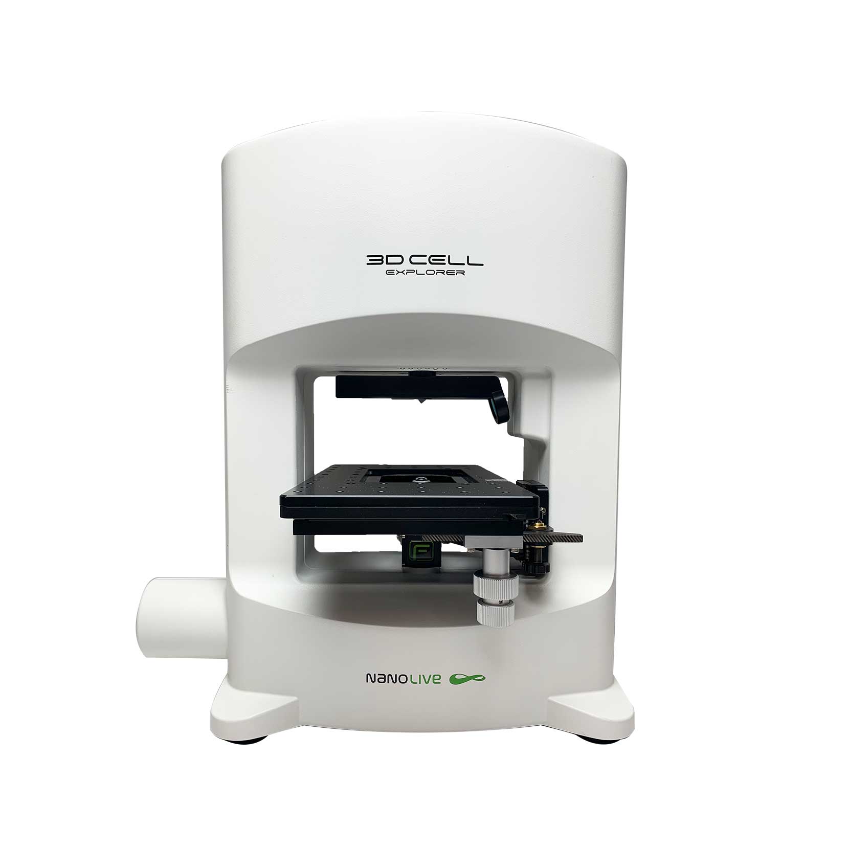
3D CELL EXPLORER-fluo
Multimodal Complete Solution: combine high quality non-invasive 4D live cell imaging with fluorescence
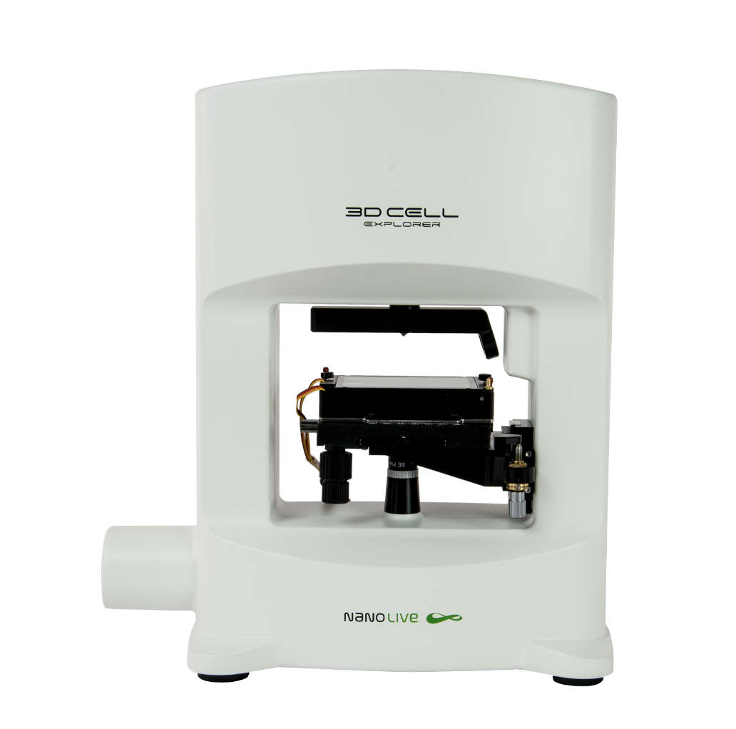
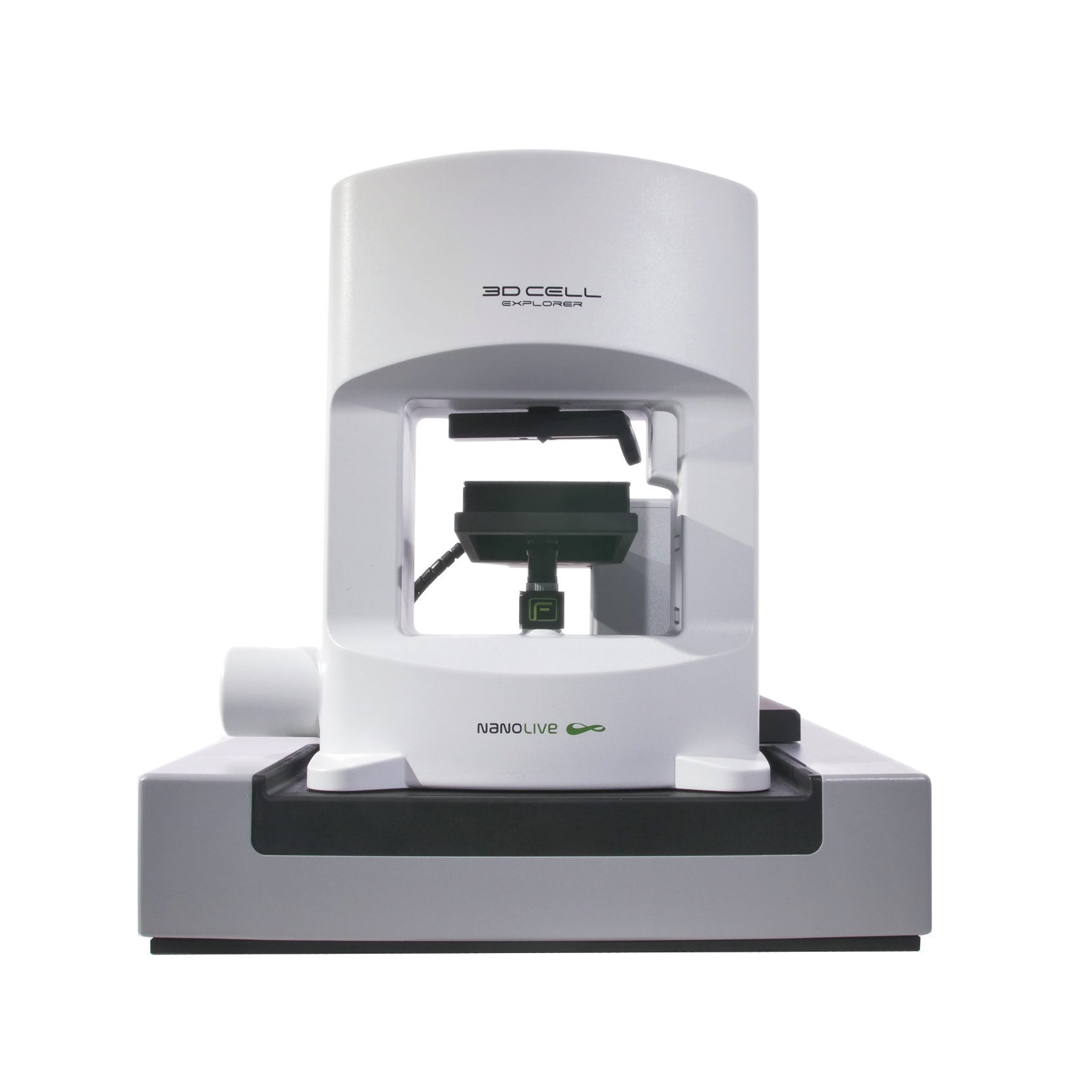
3D CELL EXPLORER
Budget-friendly, easy-to-use, compact solution for high quality non-invasive 4D live cell imaging

