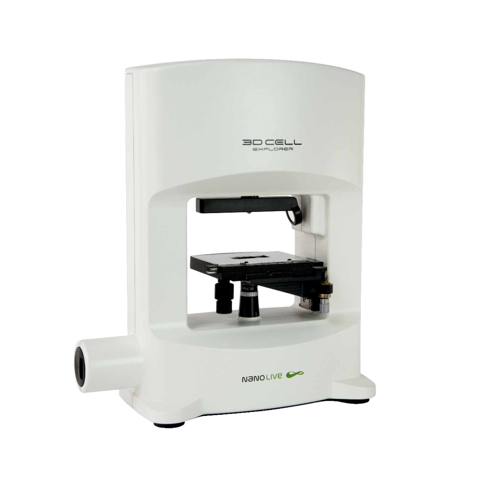
Stem cells were isolated from the umbilical cord matrix and grown in culture for 14 days. At this stage most cells in the population are at the mature megakaryocyte stage and can start producing platelets in internal membranes present in their cytoplasm. In this footage (images taken at a frequency of one image per minute), we observe the formation and spontaneous release of platelets using Nanolive’s 3D Cell Explorer.
Being able to image such processes in living megakaryocytes and without the constraints imposed by fluorescent probes, opens the door towards more complicated mechanistic studies of in vitro platelet generation.
Nanolive would like to thank the team at the Transfusion Research Center (TReC) – Belgian Red Cross-Flanders for preparing the samples.
Read our latest news
Cytotoxic Drug Development Application Note
Discover how Nanolive’s LIVE Cytotoxicity Assay transforms cytotoxic drug development through high-resolution, label-free quantification of cell health and death. Our application note explores how this advanced technology enables real-time monitoring of cell death...
Investigative Toxicology Application Note
Our groundbreaking approach offers a label-free, high-content imaging solution that transforms the way cellular health, death, and phenotypic responses are monitored and quantified. Unlike traditional cytotoxicity assays, Nanolive’s technology bypasses the limitations...
Phenotypic Cell Health and Stress Application Note
Discover the advanced capabilities of Nanolive’s LIVE Cytotoxicity Assay in an application note. This document presents a detailed exploration of how our innovative, label-free technology enables researchers to monitor phenotypic changes and detect cell stress...
Nanolive microscopes

3D CELL EXPLORER
Budget-friendly, easy-to-use, compact solution for high quality non-invasive 4D live cell imaging

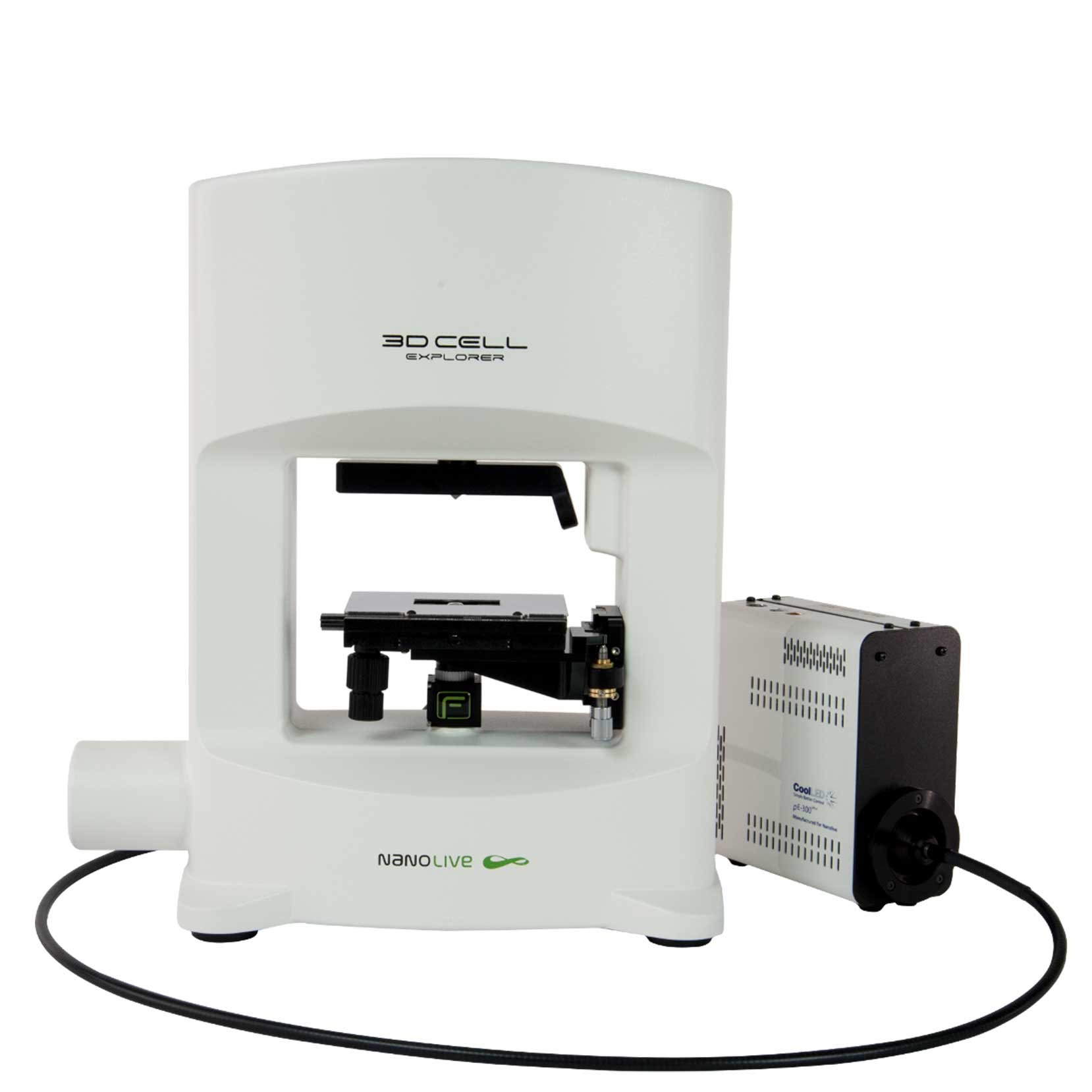
3D CELL EXPLORER-fluo
Multimodal Complete Solution: combine high quality non-invasive 4D live cell imaging with fluorescence

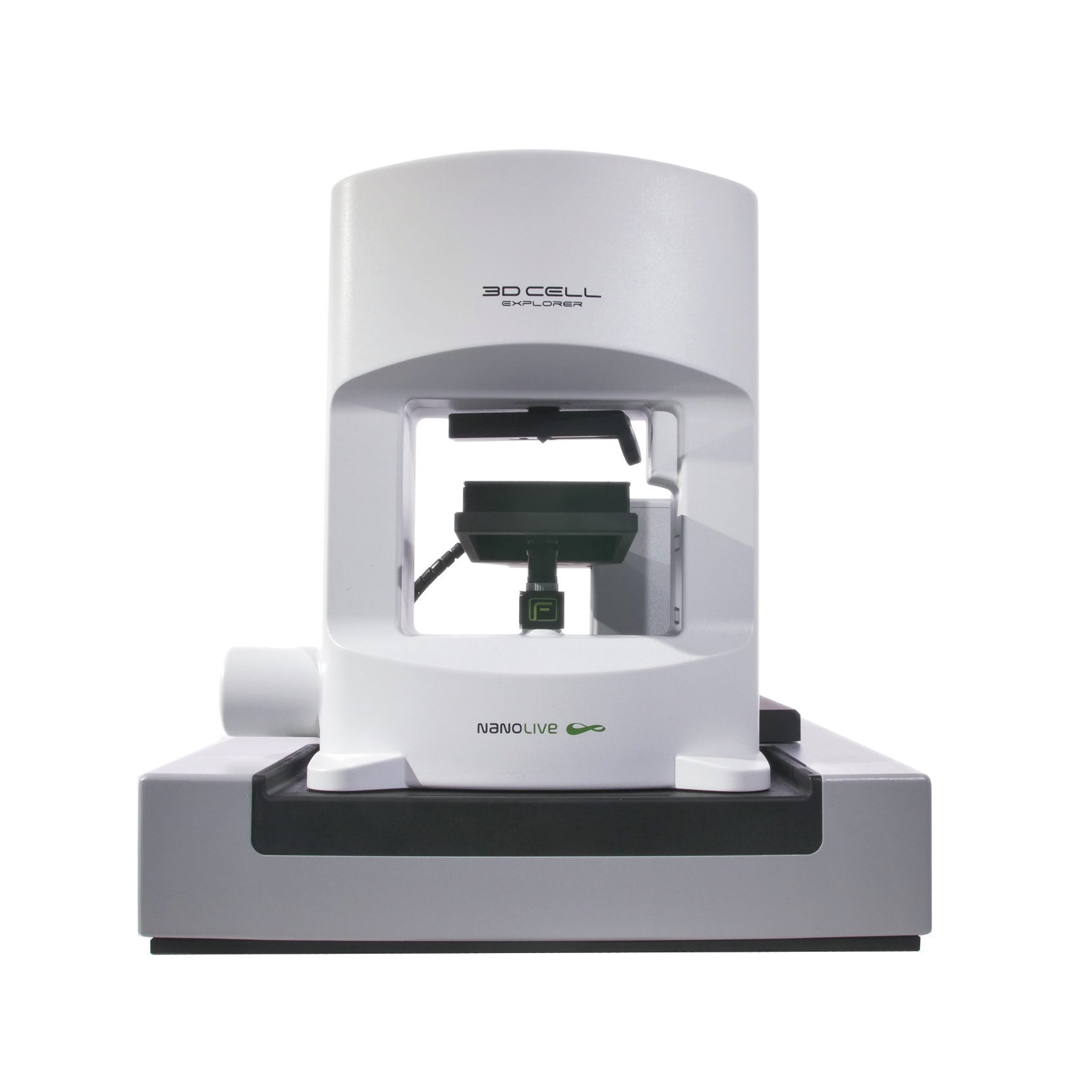
CX-A
Automated live cell imaging: a unique walk-away solution for long-term live cell imaging of single cells and cell populations


