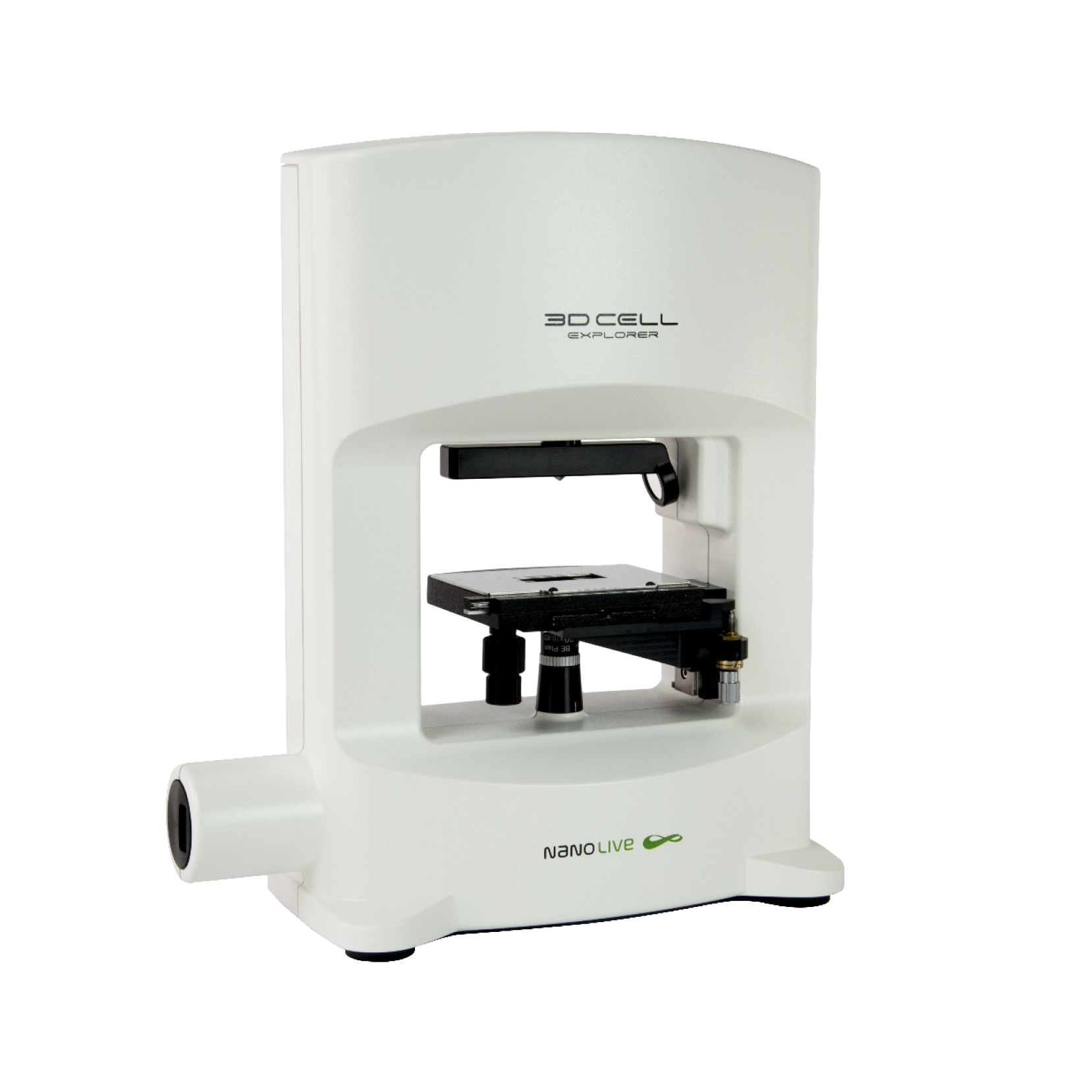
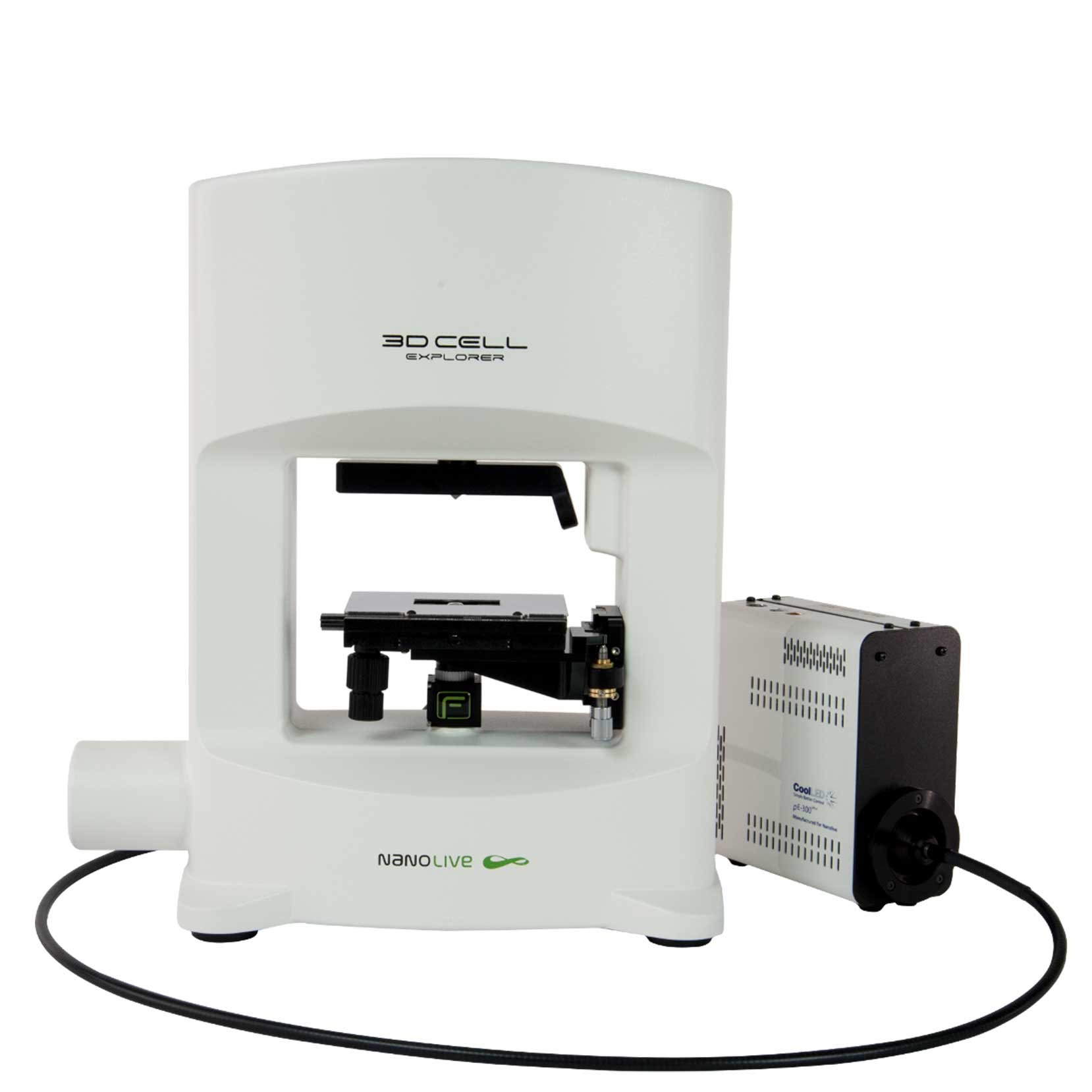
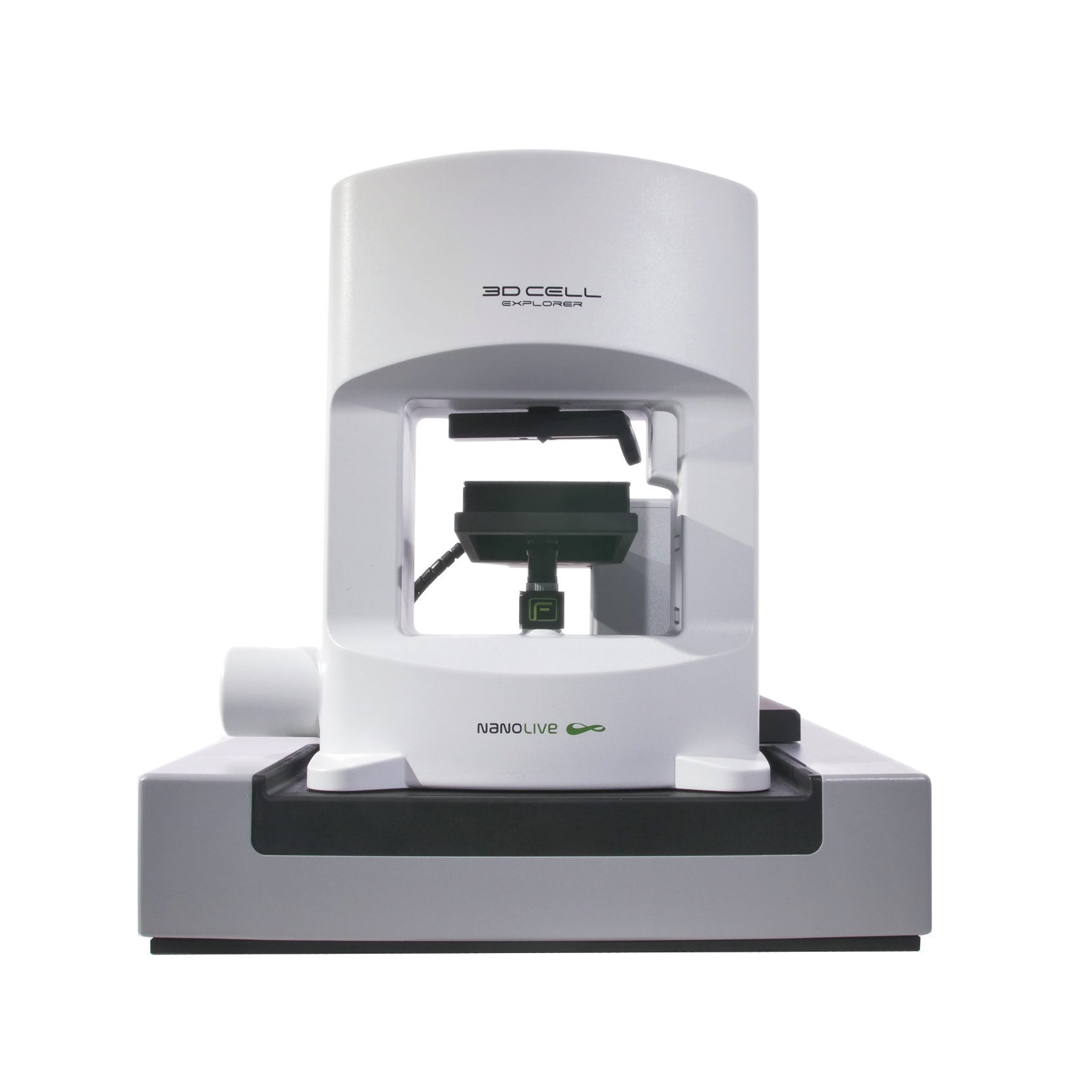
Dr. Michal Cifra is the young, dynamic team leader of the Bioelectrodynamics team at the Institute of Photonics and Electronics Academy of Sciences (Czech Republic). His lab has recently purchased Nanolive’s 3D Cell Explorer-fluo microscope and published his first paper using it in the scientific Journal of Photochemistry and Photobiology B: Biology (see here). Dr. Cifra was kind enough to meet with us to discuss the focus of his lab, the findings of his paper and give us a preview into some of the exciting plans he has in store for his new purchase.
“The current goal of my lab is to develop chip-based methods which employ electric and electromagnetic pulses to modulate the function of reconstituted cytoskeleton-based protein nanostructures outside of the cell.” The chip-based experimental systems essentially send very short (ns) electric pulses to these structures, which modulate their function and so, the response of the cell. Dr. Cifra’s lab recently demonstrated this using tubulin. The work was conducted by a former postdoc Dr.Djamel Eddine Chafai, where he showed that the electric pulses modified tubulin conformation so that it temporarily formed structures that were very different from microtubules. Moreover, the effect in cells resulted in the remodeling of the microtubule cytoskeleton. Dr. Cifra’s lab was awarded a prestigious grant worth almost €2 million over the next five years to further explore this topic using very high frequency (sub-terahertz) pulses.
We want to understand how various electromagnetic fields affect the remodeling of the cytoskeleton in real-time with all associated physiological events and Nanolive imaging is an excellent commercial solution for this. We want to use our 3D Cell Explorer-fluo to monitor morphological changes in cells in real time.
I asked Dr. Cifra how Nanolive imaging benefited his latest scientific publication, which was conducted in collaboration with scientists from the Shahid Beheshti University of Medical Sciences (Tehran, Iran). There, Dr. Djamel Eddine Chafai with his trainee Hadi Sardarabadi from Iran were instrumental in carrying out this research. “Another part of our research is dedicated to understanding biological autofluorescence (BAL) and how we can modulate it physically”. The main sources of BAL are reactive oxygen species (ROS), generated by mitochondria. The strength of the BAL signal is thus likely linked to the state of the health of a living cell/tissue/system. Detecting and measuring the signal, however, is difficult, especially when the signal strength is low. “In this paper we show that one way to boost the signal is to use mitochondria-targeted nanoparticles”. The 3D aspect of the Cell Explorer-fluo was used to determine the nanocarriers were internalized and localised in the mitochondria within the cell, and that they had no impact on the structure or morphology of the cell.
“Going 3D” Dr. Cifra advises me, is a particularly hot topic in applied physics at the moment. Scientists are extremely interested in understanding the behavior of active matter, such as microtubules. I ask him whether he has an experiment in mind. “It would be very interesting to study the polymerization and self-organization of microtubules bundles, which should contain enough matter to generate a nice refractive index signal using Nanolive imaging”. I ask whether this research has any applied relevance. “Of course, one of our end goals is to develop new electromagnetic nanobiotechnological tools that can be used to complement standard chemotherapy treatment. Most anti-cancer drugs target microtubules and so our technology could either increase the efficiency of the anti-cancer drugs or reduce the dosage of drug that is required.”
Nanolive would like to wish Dr. Cifra and his lab luck with their future research and to thank him for taking the time to talk to us.
Read our latest news
Cytotoxic Drug Development Application Note
Discover how Nanolive’s LIVE Cytotoxicity Assay transforms cytotoxic drug development through high-resolution, label-free quantification of cell health and death. Our application note explores how this advanced technology enables real-time monitoring of cell death...
Investigative Toxicology Application Note
Our groundbreaking approach offers a label-free, high-content imaging solution that transforms the way cellular health, death, and phenotypic responses are monitored and quantified. Unlike traditional cytotoxicity assays, Nanolive’s technology bypasses the limitations...
Phenotypic Cell Health and Stress Application Note
Discover the advanced capabilities of Nanolive’s LIVE Cytotoxicity Assay in an application note. This document presents a detailed exploration of how our innovative, label-free technology enables researchers to monitor phenotypic changes and detect cell stress...
Nanolive microscopes

3D CELL EXPLORER
Budget-friendly, easy-to-use, compact solution for high quality non-invasive 4D live cell imaging

3D CELL EXPLORER-fluo
Multimodal Complete Solution: combine high quality non-invasive 4D live cell imaging with fluorescence

CX-A
Automated live cell imaging: a unique walk-away solution for long-term live cell imaging of single cells and cell populations


