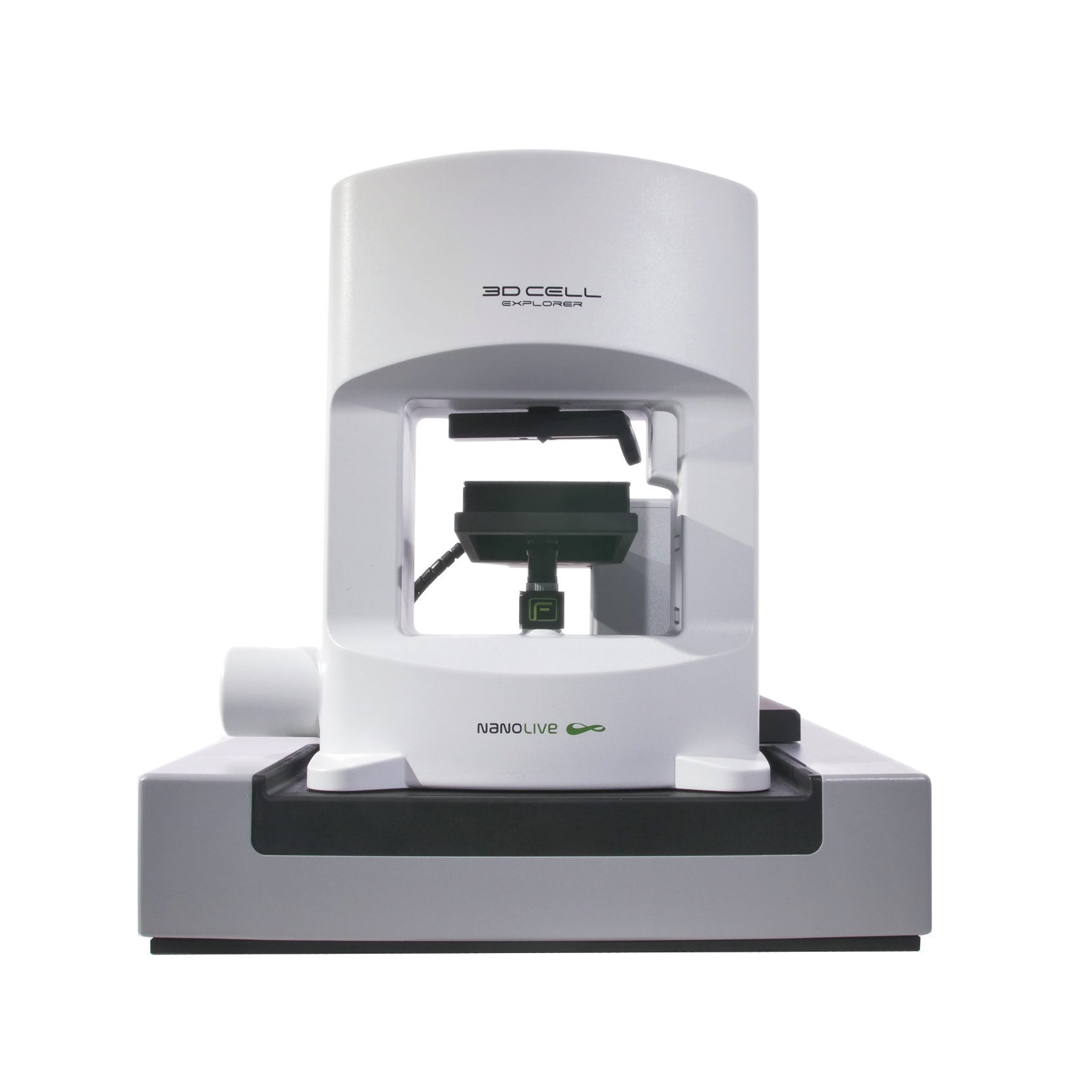
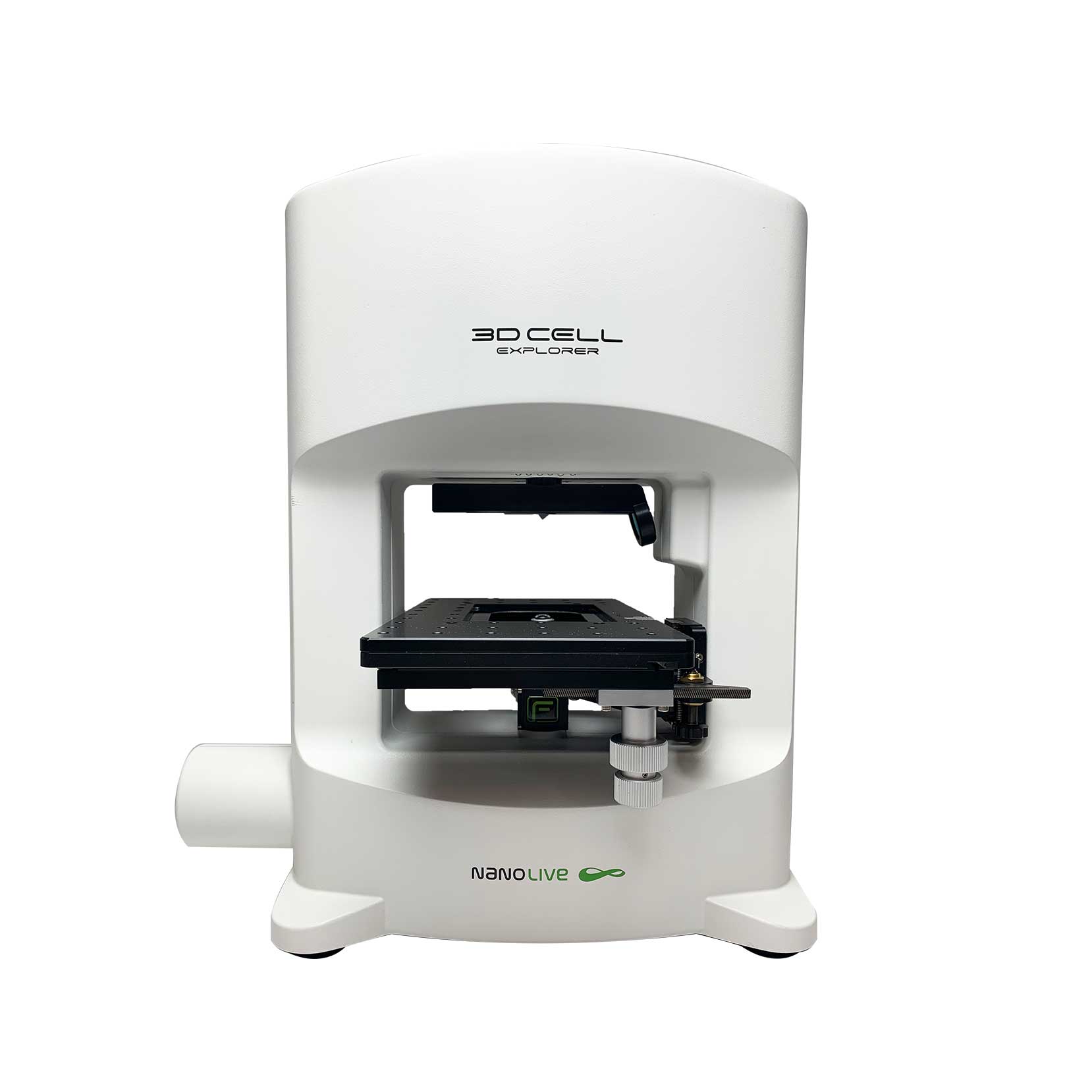
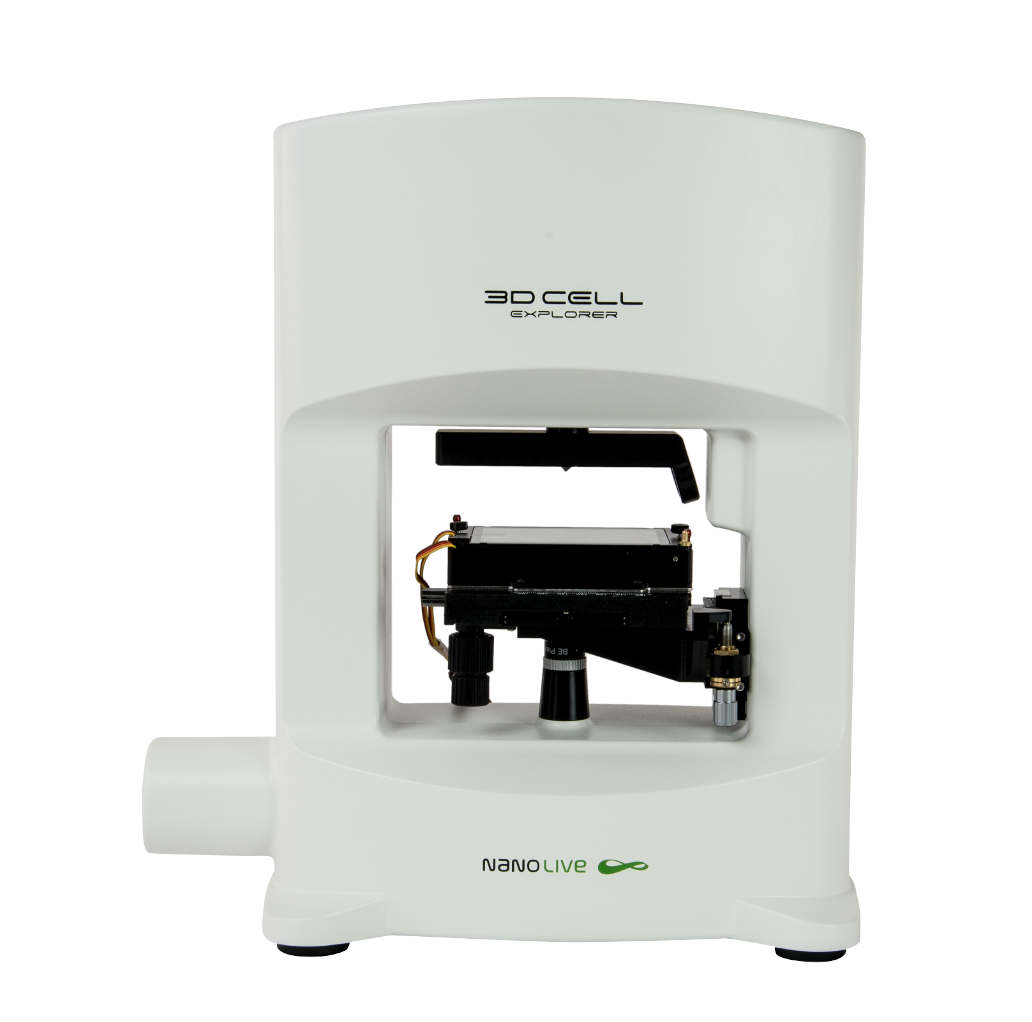
Dr. Joanna Depciuch, who runs a lab in the Department of Functional Nanomaterials, based at the Institute of Nuclear Physics Polish Academy of Sciences (Krakow, Poland), is the proud owner of a CX-A. Joanna specializes in the synthesis of nanoparticles (NPs), particularly gold NPs (AuNPs), which hold great potential for a wide range of biomedical applications including diagnostics, drug delivery, and cancer treatment (1,2). Joanna kindly agreed to meet us to explain how she plans on using her new device in her research.
AuNPs are indeed, highly flexible. By modifying the synthesis time, it is possible to alter the physical, chemical, and optical properties of the particles (3). Joanna is currently running long-term experiments to examine how cells respond to spherical, rod, and star shaped AuNPs. The video on the right, shows the morphological changes glioblastoma cancer cells undergo after the administration of gold nanorods. Images were captured every 5 mins for 4 h using the 3×3 gridscan mode on the CX-A (a field of view of 236×236 μm). Cell health is clearly negatively impacted by the presence of the AuNPs; they develop a substantial number of apoptotic bodies, and lose focal adhesion contact points, before shrinking in size.
“When you work with NPs it is essential to characterize: the number of NPs that enter the cells, the site of accumulation and the morphological response of the cells over time. The CX-A is very nice for this as we can quantify how a cell population responds to NPs with different shapes and sizes, while retaining high single-cell resolution”.
These responses are important, as they could prevent the agglomeration of cancer cells, a key process in metastasis (4, 5). So, which shape is the most successful? “Star-shaped AuNPs by far; morphological changes are observed after just 30 mins, 6 times faster than spherical AuNPs, which take 3 hours to see an effect on cell health”. The next thing Joanna plans on testing is how the type of media influences the temporal dynamics of NP accumulation. “I noticed, that if NPs are added to cell culture media, then morphological changes in the cells occur 15 mins earlier compared to if they are in water”. The idea is to optimize every step of the process, so cancer cells do not have time to aggregate.
AuNPs have another benefit over NPs made from other properties; they can be activated using 808-nm near-infrared (NIR) light to generate heat for photothermal therapy (PTT; 6-8). This is an emerging strategy for ablating solid tumours, and one that Joanna is eager to test using the fluorescence module on the CX-A. “Yes, we definitely plan on comparing whether activating AuNPs influences cell responses; it’s a very exciting field of research”. We await the results with great enthusiasm and thank Dr. Depciuch for meeting us. All the best in the future Joanna!
References
(1) Xia Y. Nat. Mat. 7(10): 758-760 (2008).
(2) Subbiah R. et al. Curr. Med. Chem. 17(36): 4559-4577 (2010).
(3) Depciuch J et al. Photodiagnosis Photodyn. Ther. 1(30): 101670 (2020).
(4) Zhao Y. et al. Adv. Drug Deliv. Rev. 30; 65(13-14): 1763-1783 (2013).
(5) Wu Y. et al. ACS Nano. 17; 12(9): 9279-9290 (2018).
(6) Lakhani PM. et al. Nanotechnology. 8; 26(43): 432001 (2015).
(7) Ali MR. et al. Proc. Natl. Acad. Sci. 11; 114(28): 5655-5663 (2017).
(8) Depciuch J et al. J. Organomet. Chem. 34 (3): e5401 (2020).
Read our latest news
Cytotoxic Drug Development Application Note
Discover how Nanolive’s LIVE Cytotoxicity Assay transforms cytotoxic drug development through high-resolution, label-free quantification of cell health and death. Our application note explores how this advanced technology enables real-time monitoring of cell death...
Investigative Toxicology Application Note
Our groundbreaking approach offers a label-free, high-content imaging solution that transforms the way cellular health, death, and phenotypic responses are monitored and quantified. Unlike traditional cytotoxicity assays, Nanolive’s technology bypasses the limitations...
Phenotypic Cell Health and Stress Application Note
Discover the advanced capabilities of Nanolive’s LIVE Cytotoxicity Assay in an application note. This document presents a detailed exploration of how our innovative, label-free technology enables researchers to monitor phenotypic changes and detect cell stress...
Nanolive microscopes

CX-A
Automated live cell imaging: a unique walk-away solution for long-term live cell imaging of single cells and cell populations

3D CELL EXPLORER-fluo
Multimodal Complete Solution: combine high quality non-invasive 4D live cell imaging with fluorescence

3D CELL EXPLORER
Budget-friendly, easy-to-use, compact solution for high quality non-invasive 4D live cell imaging


