Nanolive
Nanolive delivers breakthrough imaging solutions that combine screening, imaging, and analysis to radically advance how scientists study living cells and provide novel biological insights into the mechanisms of cancer and neurodegenerative diseases.
Our unique imaging platforms allow scientists to explore living cells in 3D without damaging them. By delivering complete measurements and understandings on a sub-cellular scale (e.g., mitochondrial networks, stem cell differentiation), our approach has the potential to accelerate research in growth industries such as drug discovery and cell therapy. But we are more than just a company, we are a family, and this is our story.
2007. An important discovery.
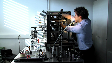
In 2007, a young and talented physicist (our future founder and CEO) Yann Cotte enrolled into a PhD program at EPFL. Yann joined the Microsystems laboratory of Prof. Christian Depeursinge, where he and another young researcher, Fatih Toy, worked on designing a device that combines holographic microscopy and computational image processing. Their goal was to build an imaging system capable of exploring living cells in 3D, without damaging them and they achieved this with stunning results. The secret to their success was to spin a low-intensity laser 360˚ around a sample taking photos and then deconvolute the images using a complex image processing algorithm to eliminate noise.
2012. A pioneering publication.
In 2012, Yann and colleagues published their ground-breaking results in the prestigious journal Nature Photonics (read here). The publication, which has currently been cited over 500 times, shook the microscopy world. An advance likened to the leap from photography to live television, the state-of-the art label-free technique puts an end to phototoxicity, making it possible to continuously image cells in a non-perturbed state for infinite periods of time.
2013. The birth of Nanolive.
In 2013, Yann met Dr. Sebastien Equis and the two founded Nanolive SA. The company, which was based at EPFL’s Innovation Park, was initially comprised of eleven people from diverse scientific and geographical backgrounds, whose goal was to develop a reliable and user-friendly commercial device based equipped with the highest quality imaging standards.

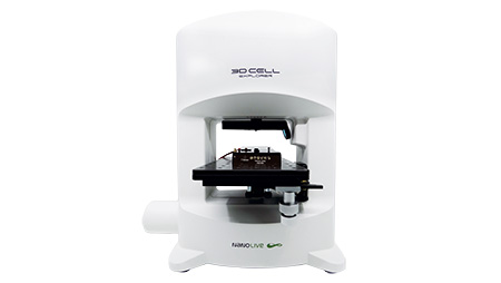
2015. The creation of the 3D Cell Explorer.
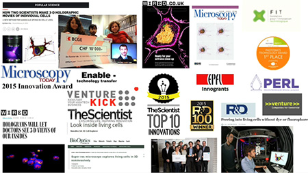
December 2015 saw the release of the highly anticipated 3D Cell Explorer. The official launch event was conducted at the annual American Society for Cell Biology conference; one of the biggest cell biology conferences in the world. Interest was high in the product, which won several prestigious awards in its first year, including the Top 10 Innovation of 2015 from The Scientist and the R&D100.
2017. The release of the 3D Cell Explorer-fluo.
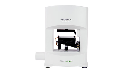
After the success of the 3D Cell Explorer, Nanolive released the World’s first holotomographic fluorescent microscope, the 3D Cell Explorer-fluo. This new microscope made it possible for researchers to combine non-invasive cell tomography with an established and recognized method: multi-channel fluorescence microscopy. Correlative imaging made it possible to test whether cell organelles such as lipid droplets and mitochondria had specific refractive indices, leading to several important proof-of-concept studies (e.g., Sandoz et al. 2019). It also allowed researchers to visualize proteins, ions, and viruses, which are otherwise too small to be captured by label-free imaging alone.
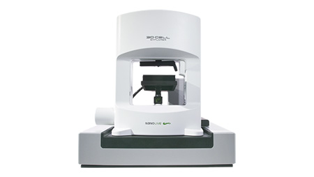
2019. The CX-A is launched
2019 saw the launch of the CX-A; the world’s first automated 96-well plate compatible label-free microscope. Automating the acquisition process while maintaining perfect device calibration and imaging focus was no mean feat and required the development of a pioneering autofocus method (explained here). The development of a stable autofocus method turned out to be a game-changer, opening a world of opportunities for microscopic screening and grid scanning (the acquisition of large fields of view). Finally, the world had a walk away solution for long-term live cell imaging that worked at scale.
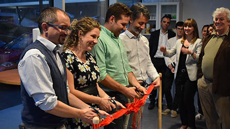
2020. New HQ opened in Tolochenaz.
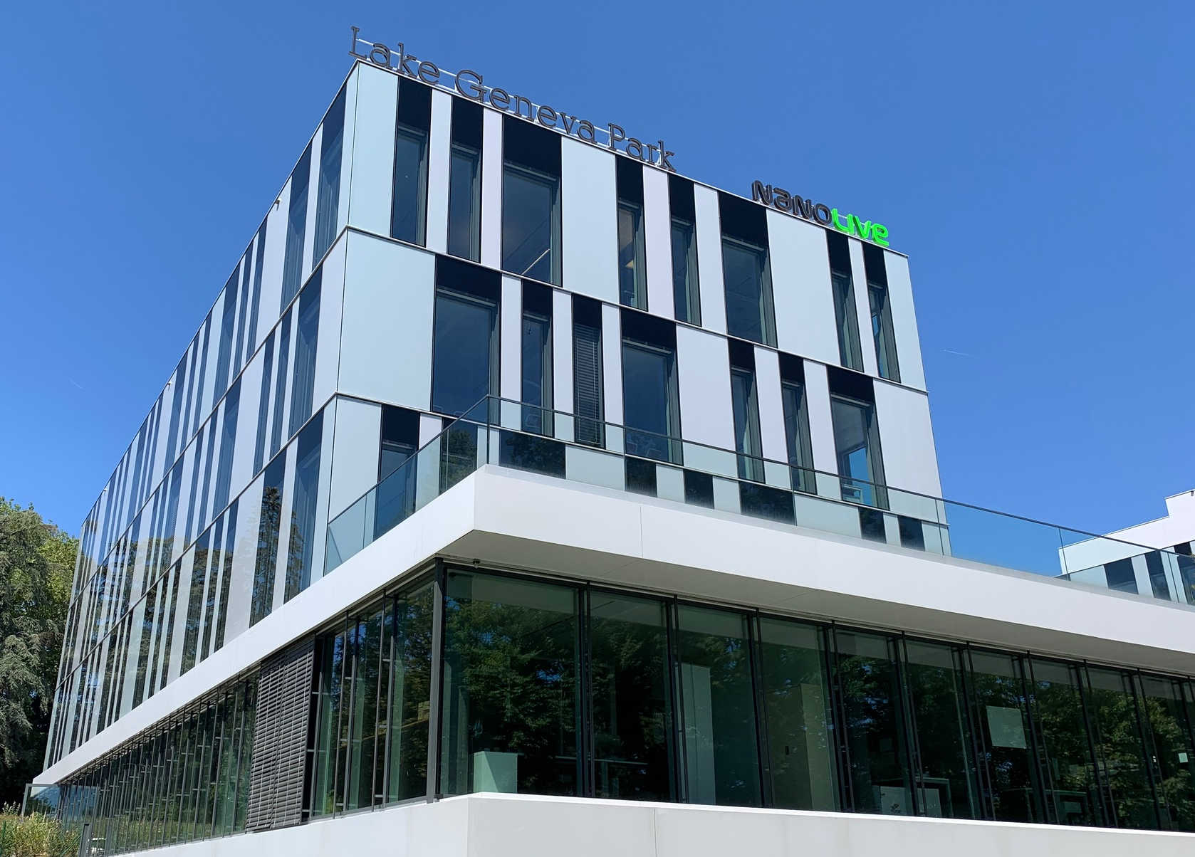
January 2020 saw a big move for Nanolive, from a very cosy office in Innovation Park to a mansion (1100 m2!) just up the road in Tolochenaz. The move was sorely needed as the Nanolive family had expanded significantly since the formation of the company, now employing more than 40 people. The building is light, open, and spacious inside, an inspiring place to work. And, even better, there is a roof-top terrace with a fantastic view overlooking Lac Leman, the perfect place to eat your lunch in Summer!
2021. EVE Analytics is unveiled.
May 2021 saw the culmination of our very own analysis program, EVE Analytics (EA). Segmenting and analyzing unstained cells that have complex textures and signals that are orders of magnitude more variable than fluorescent signals is not easy, and we are all very proud of the final product. EA quantifies 13 metrics simultaneously, 3 of which (granularity, total dry mass, and dry mass density) are unique to label-free imaging. Such high-content data allows researchers to quantify meaningful metrics with the highest biological relevance, at both the single cell and population level.
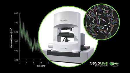
2021. The Live T Cell Assay: a revolution for immunotherapy
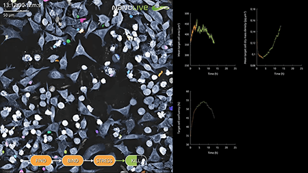
We were extremely busy in 2021 it seems as September saw the release of the Live T Cell Assay; the first software ever capable of segmenting two label-free cell populations in co-culture. The delivery of this new digital assay promises to take the analysis of cancer/T cell interactions to a whole new level. The assay allows researchers to dissect the entire T cell response, detecting the moment T cells find, bind, stress, and kill their target cells with high precision. This product holds so much potential for biotech’s involved in immunotherapy, we are impatient to see how it is received by the market.
The future. Innovation, innovation, innovation!
Our images contain such a wealth of data spanning population, single cell, and sub-cellular levels, that the future lies in developing analytical applications to harvest that data in a meaningful and simple way. This challenge will require the integration of deep learning and Artificial Intelligence (AI) into image analysis. If we do that, the therapeutic potential of our technology will really be limitless.
Read our latest news
Cytotoxic Drug Development Application Note
Discover how Nanolive’s LIVE Cytotoxicity Assay transforms cytotoxic drug development through high-resolution, label-free quantification of cell health and death. Our application note explores how this advanced technology enables real-time monitoring of cell death...
Investigative Toxicology Application Note
Our groundbreaking approach offers a label-free, high-content imaging solution that transforms the way cellular health, death, and phenotypic responses are monitored and quantified. Unlike traditional cytotoxicity assays, Nanolive’s technology bypasses the limitations...
Phenotypic Cell Health and Stress Application Note
Discover the advanced capabilities of Nanolive’s LIVE Cytotoxicity Assay in an application note. This document presents a detailed exploration of how our innovative, label-free technology enables researchers to monitor phenotypic changes and detect cell stress...


