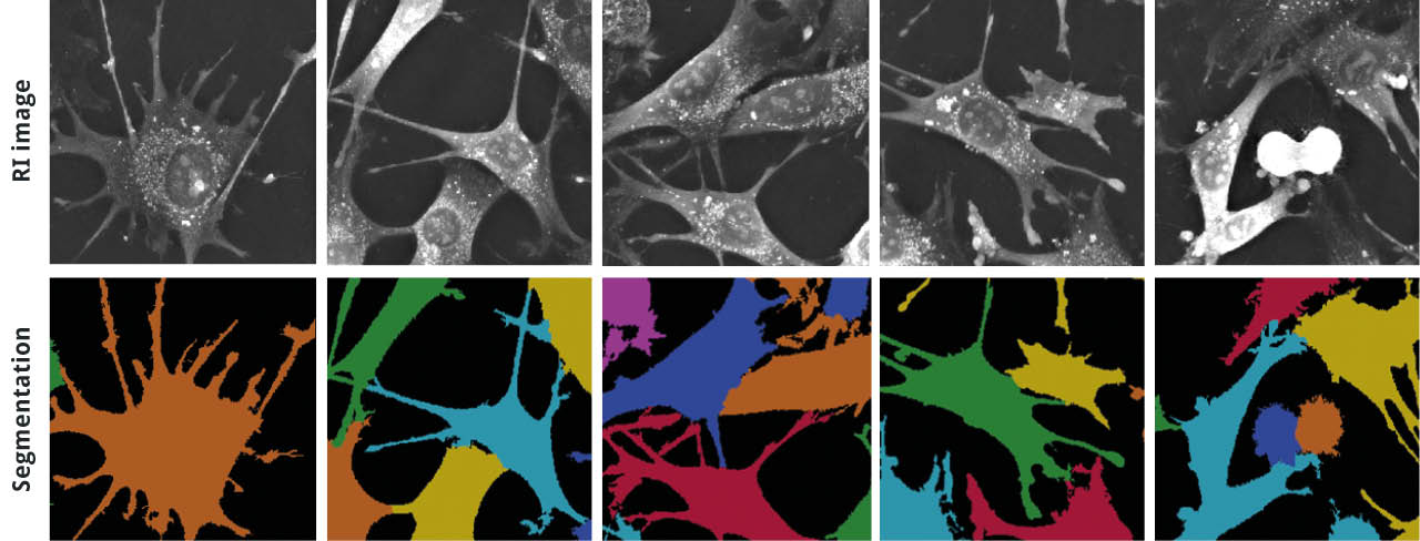
Rediscover your cells with EVE Analytics’ unique, robust, and high-precision segmentation technique
Thanks to EVE Analytics we can now offer precise cell segmentation of living cells for virtually unlimited periods of time. EVE Analytics can segment numerous single cells over thousands of images with no change in quality, regardless of the level of cell crowding/confluence in the image.
Click here to get EVE Analytics for the 3D Cell Explorer-fluo!
Applications
Quantifying changes in granularity (cell composition) during cell division
In this video, we show how granularity – a texture metric that describes a cell’s internal composition – changes during mitosis. Mesenchymal stem cells were imaged once every 15 secs for 50 mins. The cell in the top right of the video divides into two daughter cells at minute 38 in the video, which corresponds to the lowest granularity value. This example highlights the potential Nanolive imaging holds for investigating biological processes.
Quantifying the intracellular replication of pathogens inside their host cells
In this video, we quantify changing in the total dry mass that accompany the intracellular accumulation of bacteria in HeLa cells incubated with Staphylococcus aureus and imaged every 20 secs for 20 mins. Graph shows an accumulation of total dry mass as the bacteria replicate inside the cells. Images courtesy of Dr. Ana Eulalio from the University of Coimbra (Portugal).
The 3D Cell Explorer-fluo – a multimodal complete solution for 4D live cell imaging & fluorescence
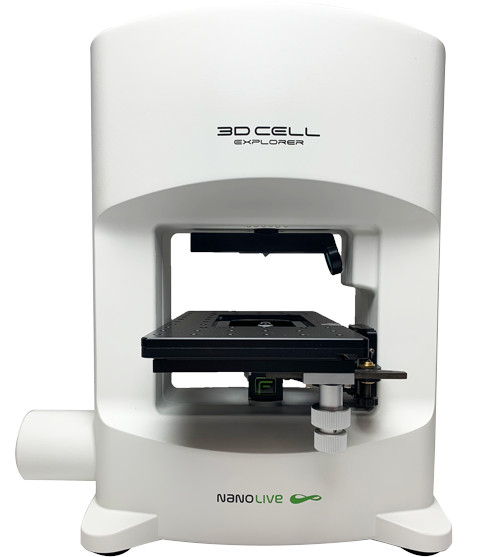
Image: 3D Cell Explorer-fluo
- Combine fully label-free imaging with correlative epifluorescence to cover a broad array of live cell imaging requirements for your lab, department, or institute
- Rediscover your living cells through multiplexing: Nanolive datasets contain the simultaneous acquisition of several biological features and organelles – therefore capturing various pathways of cell biology in real-time
- Extend Live Cell Imaging: image your live cells as long as you need. Limit cell damages caused by fluorescent markers, bleaching and phototoxicity
- Visualize the invisible: the 3D Cell Explorer-fluo allows you to visualize what would be too small to be captured label-free, such as: proteins, ions (e.g. calcium), viruses, etc
Technical Note
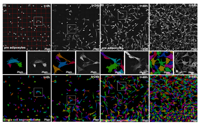
Nanolive’s label-free technology makes it possible to image cells for long periods of time, at high temporal resolution. The quantity and complexity of the images generated allows us to visualize biological processes in unprecedented detail, but also magnifies the challenges associated with image analysis. Manual image registration and analysis is impossible and so computer-aided processing must be used to harness data complexity. In this technical note, we introduce the key elements involved in cell segmentation, which are essential to understand the novelty of EVE Analytics (EA), Nanolive’s software solution for quantitative cell analysis. We then evaluate the performance of EA segmentation against fluorescence-based segmentation and compare how metrics produced by both approaches differ.
Click here to get EVE Analytics for the 3D Cell Explorer-fluo!
Read our latest news
Cytotoxic Drug Development Application Note
Discover how Nanolive’s LIVE Cytotoxicity Assay transforms cytotoxic drug development through high-resolution, label-free quantification of cell health and death. Our application note explores how this advanced technology enables real-time monitoring of cell death...
Investigative Toxicology Application Note
Our groundbreaking approach offers a label-free, high-content imaging solution that transforms the way cellular health, death, and phenotypic responses are monitored and quantified. Unlike traditional cytotoxicity assays, Nanolive’s technology bypasses the limitations...
Phenotypic Cell Health and Stress Application Note
Discover the advanced capabilities of Nanolive’s LIVE Cytotoxicity Assay in an application note. This document presents a detailed exploration of how our innovative, label-free technology enables researchers to monitor phenotypic changes and detect cell stress...
Nanolive microscopes
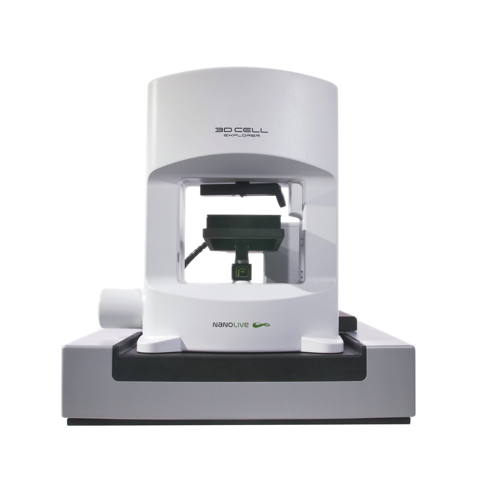
CX-A
Automated live cell imaging: a unique walk-away solution for long-term live cell imaging of single cells and cell populations
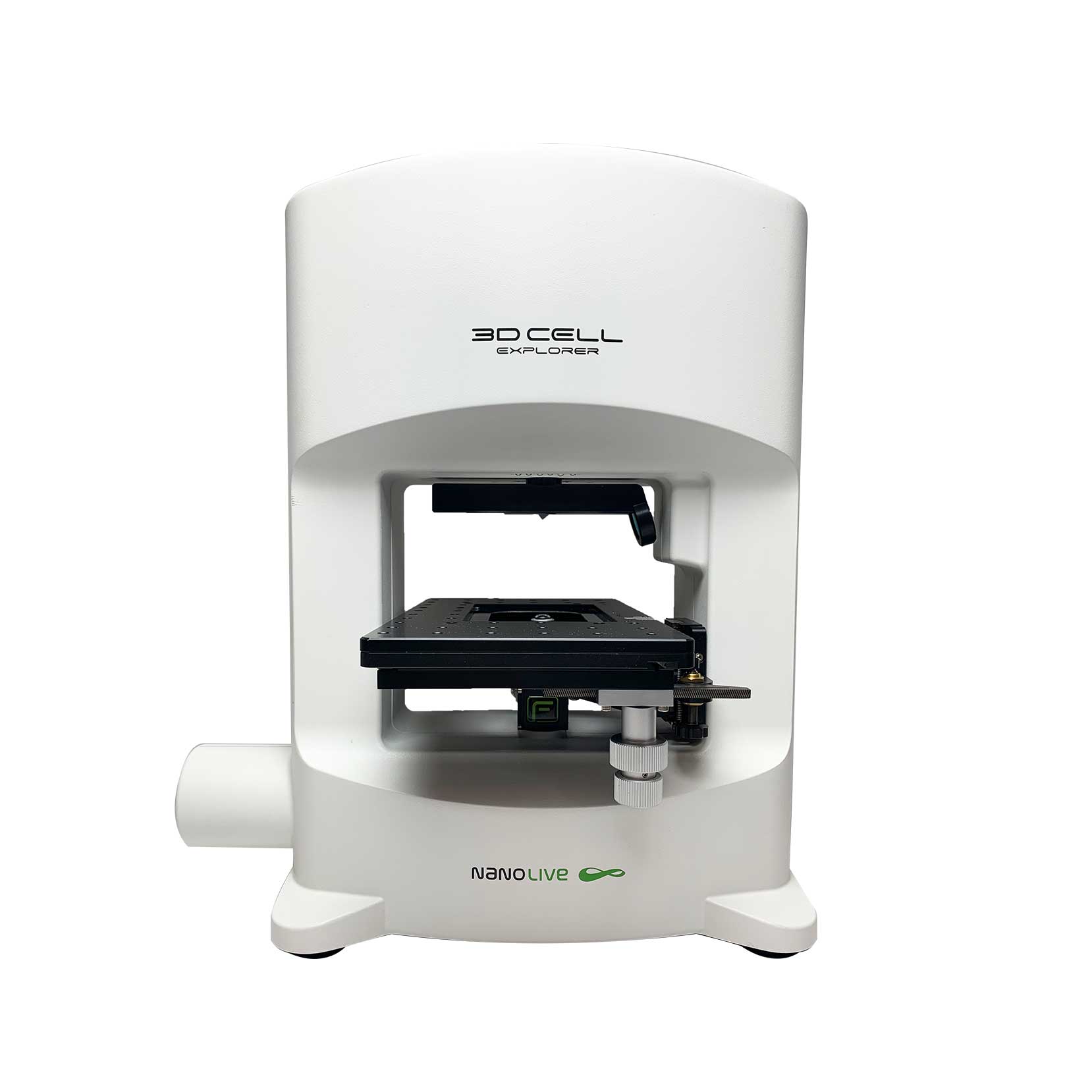
3D CELL EXPLORER-fluo
Multimodal Complete Solution: combine high quality non-invasive 4D live cell imaging with fluorescence
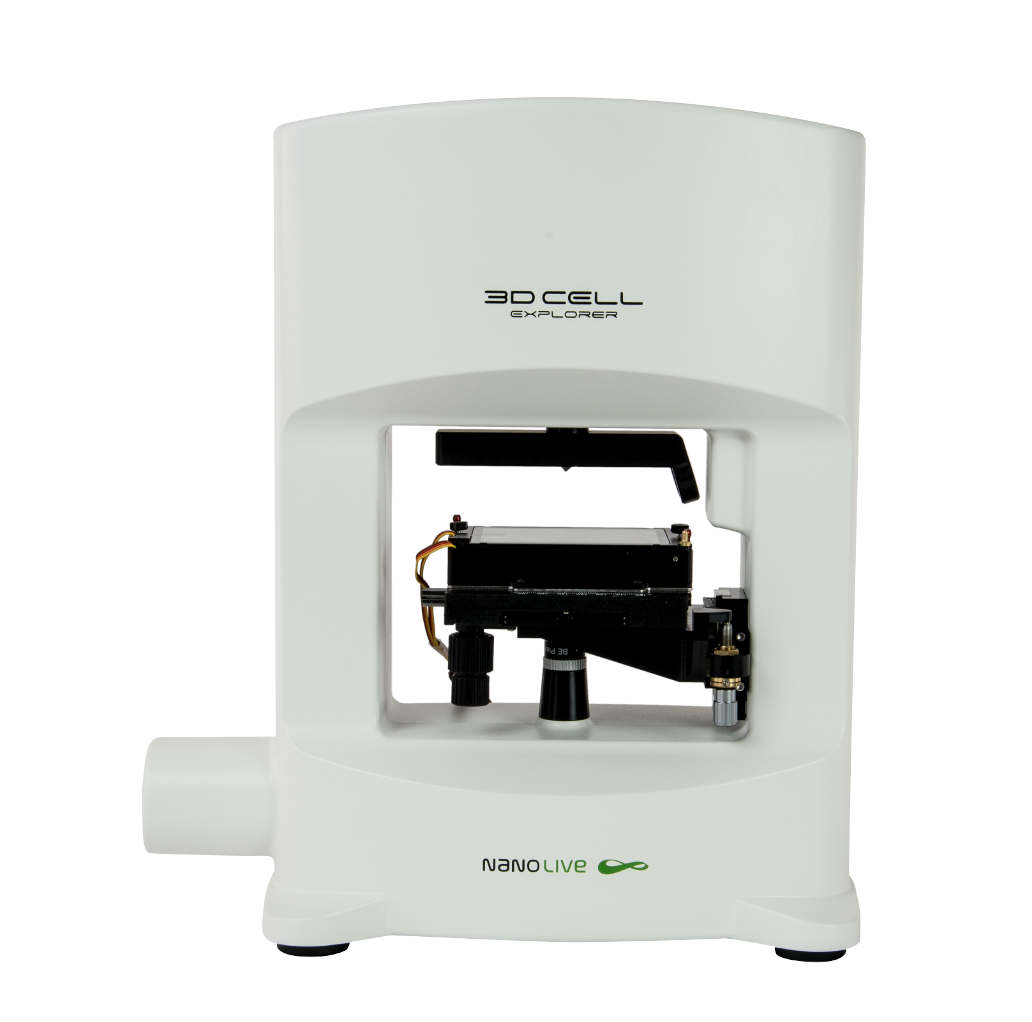
3D CELL EXPLORER
Budget-friendly, easy-to-use, compact solution for high quality non-invasive 4D live cell imaging

