Nanolive installed the CX-A automated live cell imaging platform at LUCA Science in Sapporo, Japan
Nanolive SA announced today the installation of their CX-A imaging platform at LUCA Science’s Research & Development Center in Sapporo, Japan. LUCA Science is developing a novel functional mitochondrial therapy using its proprietary mitochondria isolation technology to improve cellular bioenergetics in injured tissues. Nanolive’s label-free, live cell imaging solution advances mitochondrial research by permitting high-resolution visualization of mitochondrial dynamics with a low-intensity, non-invasive and non-phototoxic light source.
About LUCA Science
LUCA Science’s catchphrase is “Energy for Life”, and their mission is to restore the energy needed for biological processes, by using energy-generating mitochondria themselves to treat patients and improve health. Powering LUCA Science is their isolated mitochondria Q which is in development for use as an off-the-shelf therapy to treat patients with dysfunctional mitochondria in mitochondrial diseases, improving CAR-T function, reducing ischemia-reperfusion injury, and other applications.

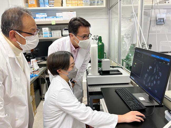
Dr. Hisashi Ohta, Head of Research and Development, Masae Takeda, Technical Staff and Ph.D. Candidate, and Dr. Rick C. Tsai, President and CEO of LUCA Science (left to right) review the first captures made with Nanolive’s CX-A platform.
Videos captured by LUCA Science
Seeing is believing: Mitochondrial uptake by cells
Here, a cell ‘reaches out’ its plasma membrane, engulfing a fluorescently-tagged mitochondrion (red). The mitochondrion is taken up and internalised by the cell.
Video captured by scientists at LUCA Science, Japan. LUCA Science has a proprietary technology to produce storable isolated mitochondria for treatment of various diseases with damaged/impaired cell function. Their mitochondria Q do not have to be encapsulated to be taken up by cells, as this video shows.
Fluorescence (TRITC channel) and refractive index images were captured every 6 minutes for 3 hours.
Mitochondrial uptake by cells
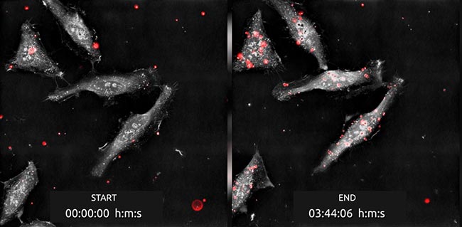
Exogenous fluorescently-tagged mitochondria Q (red) are taken up by HeLa cells over the 03 hour 44 minute imaging period.
Nanolive’s refractive index imaging allows us to see how the plasma membrane envelops mitochondria, internalizing them. 03 h 44 min after the start of imaging, many mitochondria have accumulated inside each cell.
Captured every 4 min 9 s by scientists at LUCA Science.
Visualizing endogenous mitochondria
Visualize the dynamics of this endogenous network in real time, as a cell takes in LUCA Science’s proprietary exogenous mitochondria (red) from the extracellular environment.
Captured every 7 min 40 s by scientists at LUCA Science.
Dynamic Mitosis
This HeLa cell rotated during imaging, giving us views of metaphase from the pole, and the side of the cell.
Images were captured every 1 min 23 s for 6 hours 2 minutes by scientists at LUCA Science, Japan.
“It’s very unique when you’re combining two cutting edge technologies; Nanolive’s imaging and LUCA’s Mitochondria, if one cutting edge technology shows something new, two will be even more powerful, so I hope we will be able to show many, many interesting discoveries”
Nanolive imaging helped to prove therapeutic potential
After developing a proprietary method to isolate intact and functional mitochondria, the LUCA Science team discovered that even with simple co-culture, mitochondria Q were readily taken up by the cells in vitro. Thanks to Nanolive’s live cell imaging, the uptake of mitochondria Q was captured in real-time (videos below). President and CEO of LUCA Science, Dr. Rick Tsai told us “That short video from Nanolive has converted a lot of non-believers, seeing is believing.”
Nanolive’s installation at LUCA Science
Nanolive installed the complete CX-A automated live cell imaging platform in LUCA Science’s research laboratory followed by curated training in image acquisition and analysis specific to their application in mitochondrial research. From the first acquisition, mitochondria were visible, completely label free. Combined with epifluorescence correlation, the LUCA team could easily differentiate their mitochondria Q from endogenous host cell mitochondria.

Fluorescently tagged mitochondria Q (red) are taken up by HeLa cells over the 3 h 44 min imaging period. Combining both refractive index and fluorescence imaging allows us to see how the mitochondria are able to pass through the plasma membrane, into the cells. Less than four hours after the start of imaging, many mitochondria had accumulated inside each cell.
About Nanolive in mitochondrial research
Nanolive’s live cell imaging is a go-to method for investigating mitochondria, thanks to its ability to image mitochondria as well as other organelles label-free. This non-invasive approach allows high-resolution imaging over long periods of time without damage or disruption, for a more biologically relevant picture of mitochondrial dynamics and function.
Nanolive’s Head of AI for Quantitative Biology, Dr. Mathieu Frechin said “Joining forces with LUCA Science will allow us to expand our knowledge of possible mitochondrial shapes and dynamics and possibly shed light on completely new roles for exogenous mitochondria entering cells. We are thrilled!”
Dr. Tsai summarized his hopes for the Nanolive platform at LUCA Science: “There are still many discoveries to be made on exogenous mitochondria biology, I’m convinced when we image different conditions and cell types with this platform, there will probably be valuable world-first discoveries.”
About LUCA Science Inc.
LUCA Science is a Japanese preclinical biopharmaceutical company focusing on the development of novel mitochondrial therapies to treat diseases and injuries in multiple therapeutic areas by restoring cellular bioenergetics. LUCA Science has ongoing collaborations across Japan, the US, and the UK to research the clinical applications of their mitochondrial platform.
Read our latest news
Cytotoxic Drug Development Application Note
Discover how Nanolive’s LIVE Cytotoxicity Assay transforms cytotoxic drug development through high-resolution, label-free quantification of cell health and death. Our application note explores how this advanced technology enables real-time monitoring of cell death...
Investigative Toxicology Application Note
Our groundbreaking approach offers a label-free, high-content imaging solution that transforms the way cellular health, death, and phenotypic responses are monitored and quantified. Unlike traditional cytotoxicity assays, Nanolive’s technology bypasses the limitations...
Phenotypic Cell Health and Stress Application Note
Discover the advanced capabilities of Nanolive’s LIVE Cytotoxicity Assay in an application note. This document presents a detailed exploration of how our innovative, label-free technology enables researchers to monitor phenotypic changes and detect cell stress...


