Congratulations to scientists at Seoul National University who have recently published their work in Cell Metabolism in their article “Mitochondrial fragmentation and donut formation enhance mitochondrial secretion to promote osteogenesis”! The authors characterized mitochondrial secretion by osteoblasts, and its role in regulating osteogenesis and bone regeneration1.
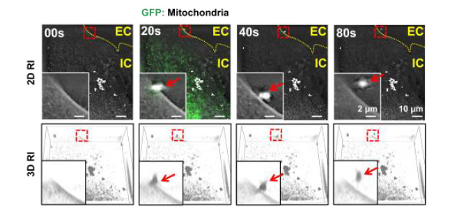
Nanolive live cell imaging was used to capture the exact moment a mitochondrion was secreted by an osteoblast (see below). Thanks to the refractive index imaging of the platform, the group were able to image cellular components including the plasma membrane label-free, reducing the amount of harmful fluorescence that cells and mitochondria were exposed to. Author Professor Lee commented “Live cell imaging using Nanolive was useful for visualizing mitochondrial secretions because it can display plasma membranes without labeling.”
Mitochondria were imaged both by refractive index and fluorescence; refractive index images were taken every 20 seconds, and fluorescence images were taken every 2 minutes (to reduce phototoxicity) for a total of 2 hours. Six refractive index images were taken for each fluorescence image, to give a higher temporal resolution of the mitochondrial dynamics. Fluorescence was used to ensure that mitochondria were accurately detected, and the group created a mouse model where osteoblast mitochondria expressed green fluorescent protein to achieve this.

Figure 1. A single GFP-tagged mitochondrion (indicated by red arrow) was captured passing through the cell membrane (light gray, imaged by refractive index). EC indicates extracellular space, IC indicates intracellular space. Taken from Figure S21.
Nanolive’s live cell imaging is a go-to method for investigating mitochondrial dynamics for many research groups, thanks to its ability to image mitochondria, as well as other organelles without labels. See the full list of publications.
References:
- Suh J. et al. Mitochondrial fragmentation and donut formation enhance mitochondrial secretion to promote osteogenesis. Cell Metabolism. 2023 Feb 7;35(2):345-360.e7. https://doi.org/10.1016/j.cmet.2023.01.003
Read our latest news
Cytotoxic Drug Development Application Note
Discover how Nanolive’s LIVE Cytotoxicity Assay transforms cytotoxic drug development through high-resolution, label-free quantification of cell health and death. Our application note explores how this advanced technology enables real-time monitoring of cell death...
Investigative Toxicology Application Note
Our groundbreaking approach offers a label-free, high-content imaging solution that transforms the way cellular health, death, and phenotypic responses are monitored and quantified. Unlike traditional cytotoxicity assays, Nanolive’s technology bypasses the limitations...
Phenotypic Cell Health and Stress Application Note
Discover the advanced capabilities of Nanolive’s LIVE Cytotoxicity Assay in an application note. This document presents a detailed exploration of how our innovative, label-free technology enables researchers to monitor phenotypic changes and detect cell stress...


