Earlier in the year we published a blogpost highlighting some of the incredible on-going research that Nanolive users in Spain and Portugal are conducting (here), and now it’s Belgium’s turn! Over the next couple of months, we will be updating this page with some of the hottest research from users based at KU Leuven and Brussels.
Dr. Sarah Borrie, Laboratory for the Research of Neurodegenerative Diseases (VIB-KU Leuven Center for Brain & Disease Research)
The last video featured in this series comes from Dr. Sarah Borrie, a post-doctoral researcher in the Laboratory for the Research of Neurodegenerative Diseases, that is headed by Prof. Bart De Strooper. Prof. De Strooper and his team study the mechanisms that are involved in neurodegenerative diseases, such as Dementia, Parkinson’s Disease and Alzheimer’s Disease (AD). Dr. Borrie focuses on the role that microglia (innate immune cells in the central nervous system) play in AD.
In the decades before the onset of dementia symptoms, extracellular amyloid-beta (Aβ) deposits (also known as amyloid plaques) accumulate in the brain and microglia amass within and around these structures. These changes in distribution and morphology of microglia have long been advocated as being important in the development of AD. Indeed, Alois Alzheimer himself noted that “the glia have developed numerous fibers” and “many glial cells show adipose saccules” in his seminal work in 1907. Yet, the exact mechanism that turns normally protective cells into a harmful presence has never been elucidated. Live cell imaging may be able to shine some light on this “switch” as it provides dynamic and kinetic information about changes in cell phenotype and function.
In this video, we observed human microglia cells stimulated by Aβ fibrils. Images were captured once every 20 secs for 12 h using Nanolive’s 3D Cell Explorer-fluo microscope.
Dr. Borrie explained some of the interesting observations she gained from the use of live cell imaging. The main thing that surprised her, was how motile these cells really are.
“Normally, we fix our cells, and we always see many rounded cells in the images. We thought the rounded morphology was related to the physiological state of the cell, but it was not clear. The video taken using the Nanolive microscope revealed that in fact, the rounded cells are highly motile microglia. This may allow us to refine how we categorise these microglia. This for me, is the real power of real time imaging.”
Another thing that impressed Dr. Borrie was the high variability of cell morphologies that exists in the population. Some cells (e.g., the cell at the bottom of the screen) are large and very static, so are amoeboid, while others are much smaller and more motile. In the future, it could be interesting to quantify the variation in morphology that exists between single cells in a population.
Dr. Emiel Michiels, VIB-KU Leuven Centre for Brain and Disease Research
Protein misfolding and deposition are common features of many neurodegenerative disorders including Alzheimer’s Disease (AD), the world’s leading cause of dementia. The Switch Laboratory, jointly headed by Prof. Frederic Rousseau and Prof. Joost Schymkowitz focuses on understanding the mechanisms that control protein folding and the role that misfolding plays in disease.
Two groups of proteins, amyloid beta and tau proteins, are important in the development and progression of neurodegenerative diseases. Abnormally folded amyloid beta proteins form extracellular amyloid plaques, while tau protein abnormalities lead to the creation of neurofibrillary tangles inside nerve cell bodies. Both reduce neuronal functioning and connectivity, causing a progressive loss of brain function.
In this video, taken by post-doctoral researcher Dr. Emiel Michiels, we observe how human embryonic kidney (HEK) cells that are engineered to report tau seeding activity (cell line: Tau RD P301S FRET Biosensor cells, ATCC CRL-3275) respond to fluorescently labeled extracellular Tau seeds (Atto-633). Refractive index images were taken every 10 secs for 1 h and 20 mins, while fluorescent images (red and green channels) were taken every 3 mins 20 secs (not all fluorescent images are shown in the video).
Red fluorescence indicates the position of extracellular tau seeds in the culture media, while green fluorescence reports when cells nucleate the aggregation of endogenous tau reporter proteins.
The video allows two very interesting observations to be made:
(1) the position of the red fluorescence inside the cell proves that extracellular tau seeds are internalized by the HEK cells and
(2) comparing the red fluorescence with the refractive index image shows that aggregations of intracellular tau produce a high enough RI signal to visualize them using RI alone. This last finding is an important proof-of-concept that Nanolive imaging can be used to investigate tau aggregation in a label-free manner in the future.
A. Prof. Lynette Lim, VIB-KU Leuven
Next, we feature a video from Assistant Professor Lynette Lim, the Group leader of the Laboratory of Interneuron Developmental Dynamics at VIB-KU Leuven Center for Brain & Disease Research, showing the behaviour of neural progenitor cells (NPCs) in culture.
Derived from neural stem cells, NPCs play a key role in the development of the mammalian cerebral cortex, the part of the brain responsible for behaviours such as perception, cognition, memory, and emotion. The Lim lab focuses on understanding the metabolic and transcriptomic processes that control neuronal diversity and circuit assembly during the development of the cerebral cortex and so, visualizing dynamic processes label-free and with single cell resolution is of interest.
In this video (images taken every 20 secs for 15 h using Nanolive’s 3D Cell Explorer-fluo), we observe NPC cell mitosis in stunning detail. NPCs are only able to divide a limited number of times and unlike neural stem cells, are unable to self-renew. This act of division may thus be the first step of differentiation towards the formation of neuronal or glial cells; cells that eventually form most of the central nervous system.
In this respect it is pertinent to consider the behaviour of the two daughter cells after their creation. Both cells develop dynamic protrusions that repeatedly pulse and coalesce; a type of behaviour that has been linked to cellular adhesion and self‐organization in other cell types using Nanolive imaging (see here).
As proliferation, migration, and organization define the key stages in cortex development, Nanolive imaging appears to be a promising avenue for future research in this field.
Dr. Ryohei Iwata, VIB-KU Leuven
Up first, is a video taken by Dr. Ryohei Iwata, a post-doctoral fellow in the Laboratory of Stem Cell and Developmental Neurobiology at VIB-KU Leuven. The lab, headed by Prof. Pierre Vanderhaeghen, aims to elucidate the key mechanisms that control the development of the cerebral cortex to understand how the human brain evolves and how genetic diseases arise.
Human pluripotent stem cell (hPSC)-derived cortical cells are popular in vitro models in neuroscience because they produce neurons with high consistency under specific differentiation conditions.
Here, we observe the dynamic behaviour of (hPSC)-derived cortical cells on Matrigel over time. Images were taken every 30 secs for 10.5 h using the 3D Cell Explorer-fluo microscope. The high spatio-temporal resolution of the images reveals how active these cells really are in culture. Elongated mitochondria-containing axonal protrusions can be observed probing the surface and interacting with their neighbouring cells, common signs of network formation.
Of particular interest is the dynamic behaviour of growth cones (zoom at 18 secs). Growth cones display a hand-like morphology. The “fingers” are made up of fine filaments called filopodia made of F-actin and contain the receptors and cell adhesion molecules required for axon growth. While the structures known as lamellipodia form the “webbing” between the filopodia. This video shows how rapidly growth cones can change direction and branch in response to changes in their microenvironment. Studying these processes is of critical importance for understanding how specified patterns of wiring between neurons are formed during development and how failures of wiring can compromise normal function.
Read our latest news
Revolutionizing lipid droplet analysis: insights from Nanolive’s Smart Lipid Droplet Assay Application Note
Introducing the Smart Lipid Droplet Assay: A breakthrough in label-free lipid droplet analysis Discover the power of Nanolive's Smart Lipid Droplet Assay (SLDA), the first smart digital assay to provide a push-button solution for analyzing lipid droplet dynamics,...
Food additives and gut health: new research from the University of Sydney
The team of Professor Wojciech Chrzanowski in the Sydney Pharmacy School at the University of Sydney have published their findings on the toxic effect of titanium nanoparticles found in food. The paper “Impact of nano-titanium dioxide extracted from food products on...
2023 scientific publications roundup
2023 has been a record year for clients using the Nanolive system in their scientific publications. The number of peer-reviewed publications has continued to increase, and there has been a real growth in groups publishing pre-prints to give a preview of their work....
Nanolive microscopes
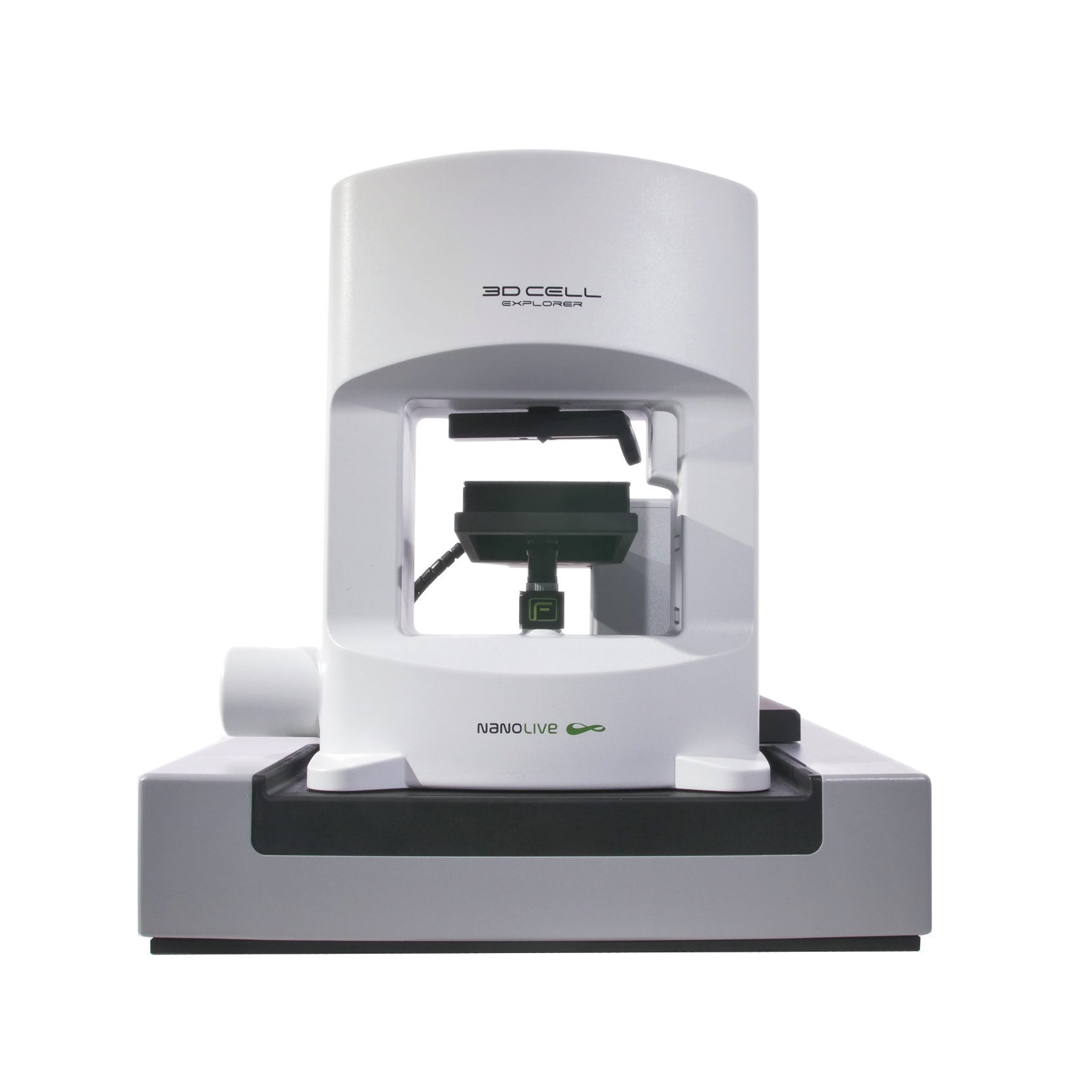
CX-A
Automated live cell imaging: a unique walk-away solution for long-term live cell imaging of single cells and cell populations
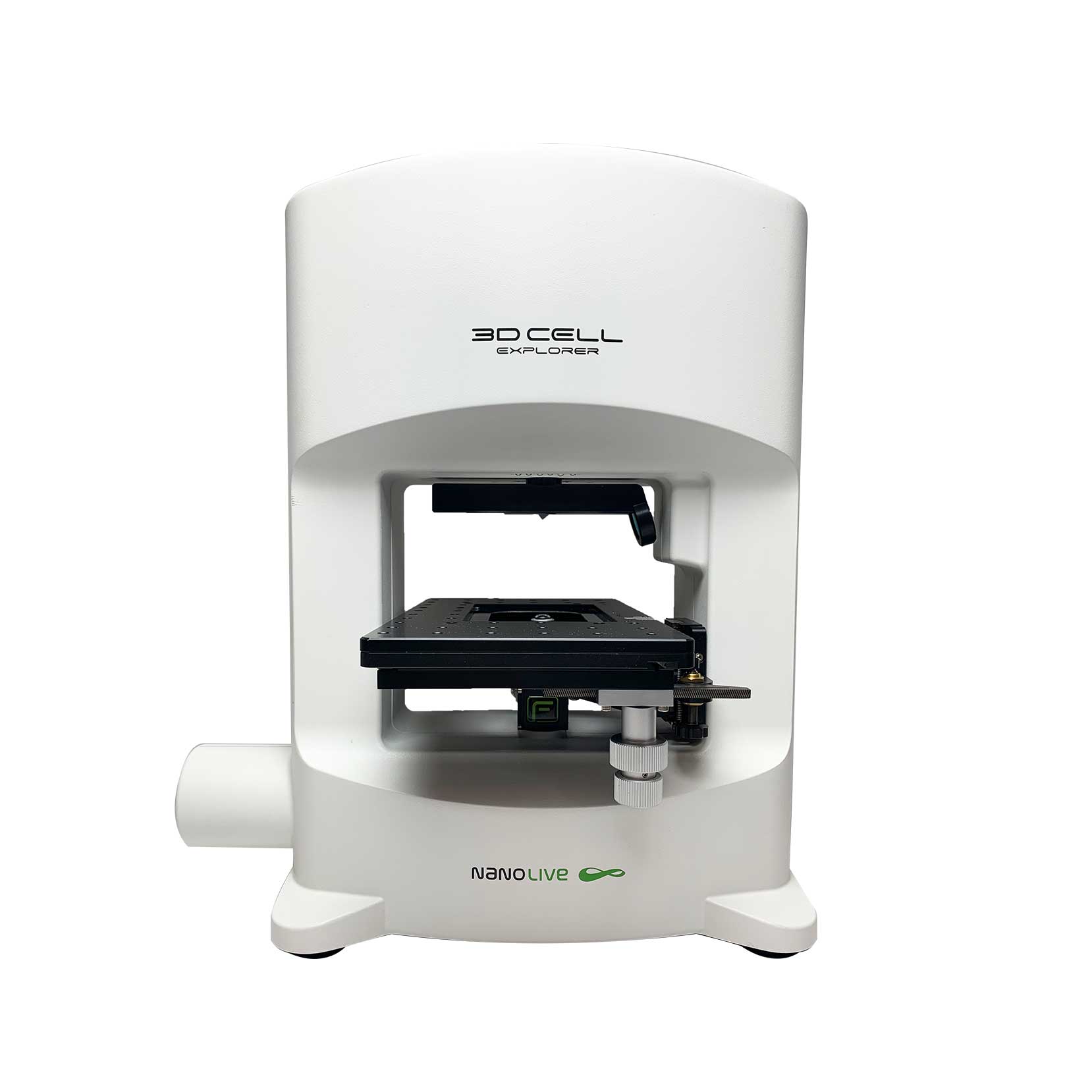
3D CELL EXPLORER-fluo
Multimodal Complete Solution: combine high quality non-invasive 4D live cell imaging with fluorescence
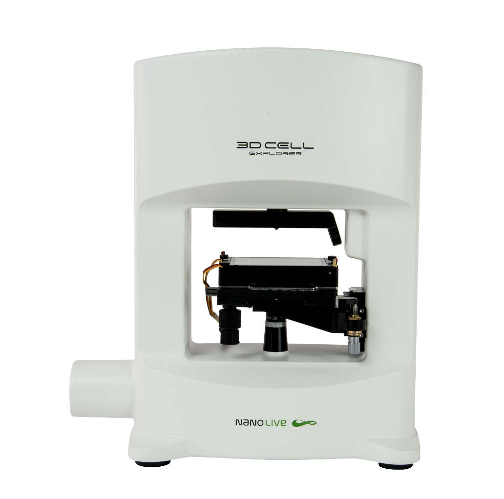
3D CELL EXPLORER
Budget-friendly, easy-to-use, compact solution for high quality non-invasive 4D live cell imaging

