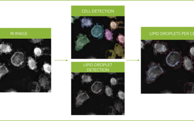
Nanolive have been working in close collaboration with swiss-based biotech firm Idorsia for several years now. Idorsia, who specialize in the discovery, development, and commercialization of innovative small molecules, have been testing our automated microscope, the CX-A and our new quantitative software, EVE Analytics. We met with Dr. Urs Lüthi the Associate Director and Deputy Head of high throughput screening (HTS), and Alexandre Peter the Principal Scientific Associate to ask them for their opinions about the Nanolive solution.
Both Urs and Alexandre were impressed by the compact design and user-friendliness of the CX-A, its environmental chamber, and the software interface. However, it was the resolution of the microscope that really blew them away.
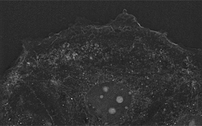
“The big advantage of Nanolive technology in my opinion, is the resolution, which we hardly get with other imaging devices. We can visualize structures, such as the nuclear envelope crystal clear and I think this is really amazing” advises Urs. Alexandre is similarly effusive, but for him, the ability to capture dynamic cell behaviours is the biggest benefit. “The videos recorded with the microscope are where you see the best of this instrument”.
The past 20 years has seen a big shift in the drug discovery process, from biochemical enzymatic assays to cell-based screens explained Urs. Live cell phenotypic screening plays an important role after the HTS stage once promising compounds are whittled down to the hundreds. This step, is where Urs sees Nanolive imaging potentially fitting in.
“Your data, with its dramatic resolution, provides a prime opportunity to measure the effect that a compound has on the morphology, pattern, and granularity of a cell, which we can use to predict the bioactivity of a compound. If we can identify compound-specific phenotypes, then we can screen other compounds for this phenotypic response and hypothesize that they function via a similar mechanism-of-action”
An application of biological fingerprints where the broadly used chemical fingerprints would fail.
This approach, however, requires high-precision cell segmentation and complete cell metrics. So, what does Alexandre think of the cell segmentations in EVE Analytics? “The quality of your segmentation is really, really good. We know that it’s not easy to segment cells in holotomographic images (due to the high texture complexity), but Nanolive is very good at it.” And the software itself? “Accessible for new users, the software guides the user well, from entering the plate parameter settings to obtaining the results.”
That is all well and good, but we want to see an example! Idorsia were kind enough to let us share the incredible results of experiment where LUHMES neuronal precursor cells were stimulated to undergo differentiation into mature dopaminergic neurons (Video 1). Cells were imaged using the 3×3 gridscan mode on the CX-A at an acquisition rate of 1 image every hour, for a total of 68 hours. By the end of the video, a mature neuronal-like network, containing extensive neurite outgrowths and growth cones is evident. And, if we zoom in, lipid droplets and mitochondria can be seen shuttling between neurons within the network.
Nanolive would like to thank Idorsia for this productive on-going collaboration. We hope our venture will result in more publications like this one.
Read our latest news
Revolutionizing lipid droplet analysis: insights from Nanolive’s Smart Lipid Droplet Assay Application Note
Introducing the Smart Lipid Droplet Assay: A breakthrough in label-free lipid droplet analysis Discover the power of Nanolive's Smart Lipid Droplet Assay (SLDA), the first smart digital assay to provide a push-button solution for analyzing lipid droplet dynamics,...
Food additives and gut health: new research from the University of Sydney
The team of Professor Wojciech Chrzanowski in the Sydney Pharmacy School at the University of Sydney have published their findings on the toxic effect of titanium nanoparticles found in food. The paper “Impact of nano-titanium dioxide extracted from food products on...
2023 scientific publications roundup
2023 has been a record year for clients using the Nanolive system in their scientific publications. The number of peer-reviewed publications has continued to increase, and there has been a real growth in groups publishing pre-prints to give a preview of their work....
Nanolive microscopes
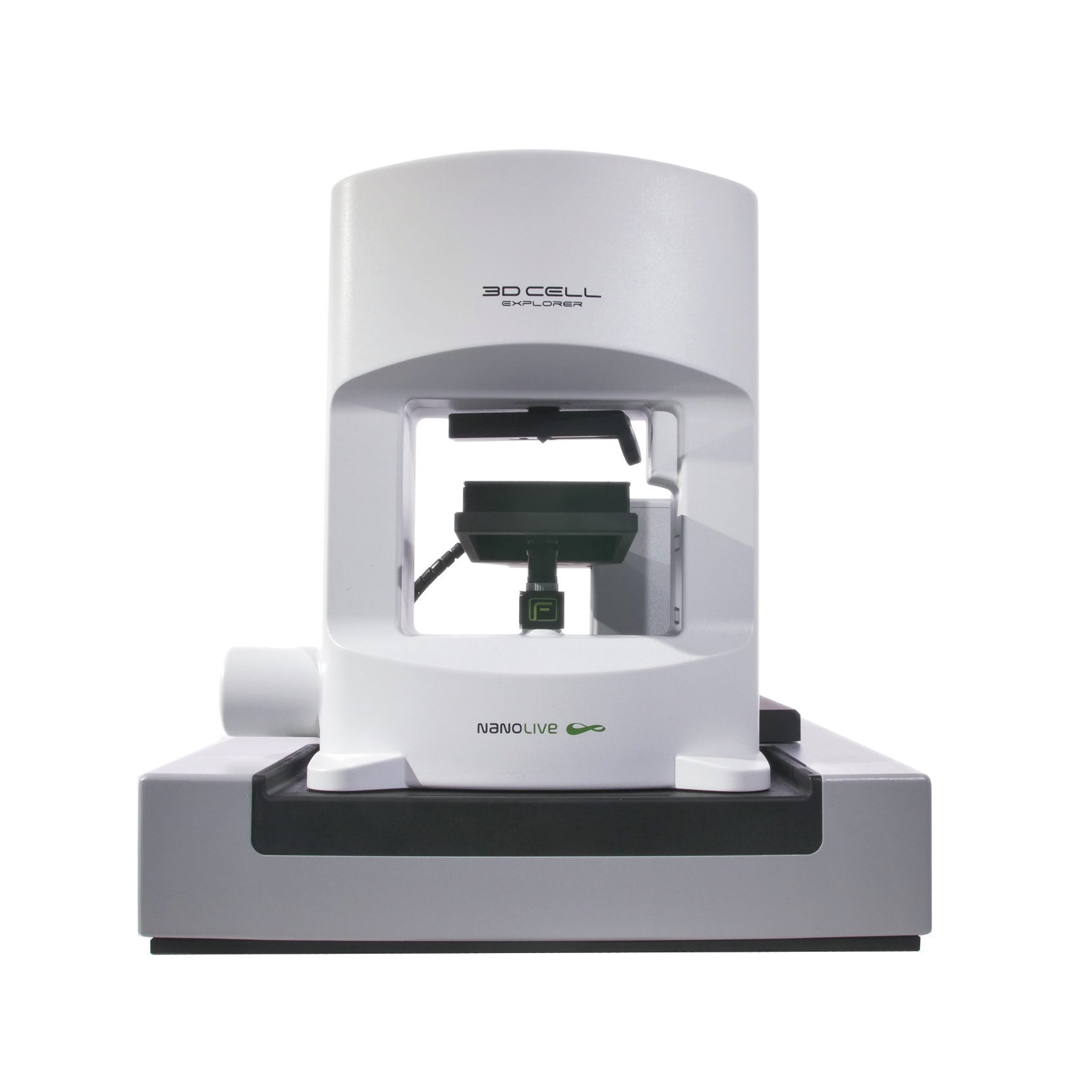
CX-A
Automated live cell imaging: a unique walk-away solution for long-term live cell imaging of single cells and cell populations
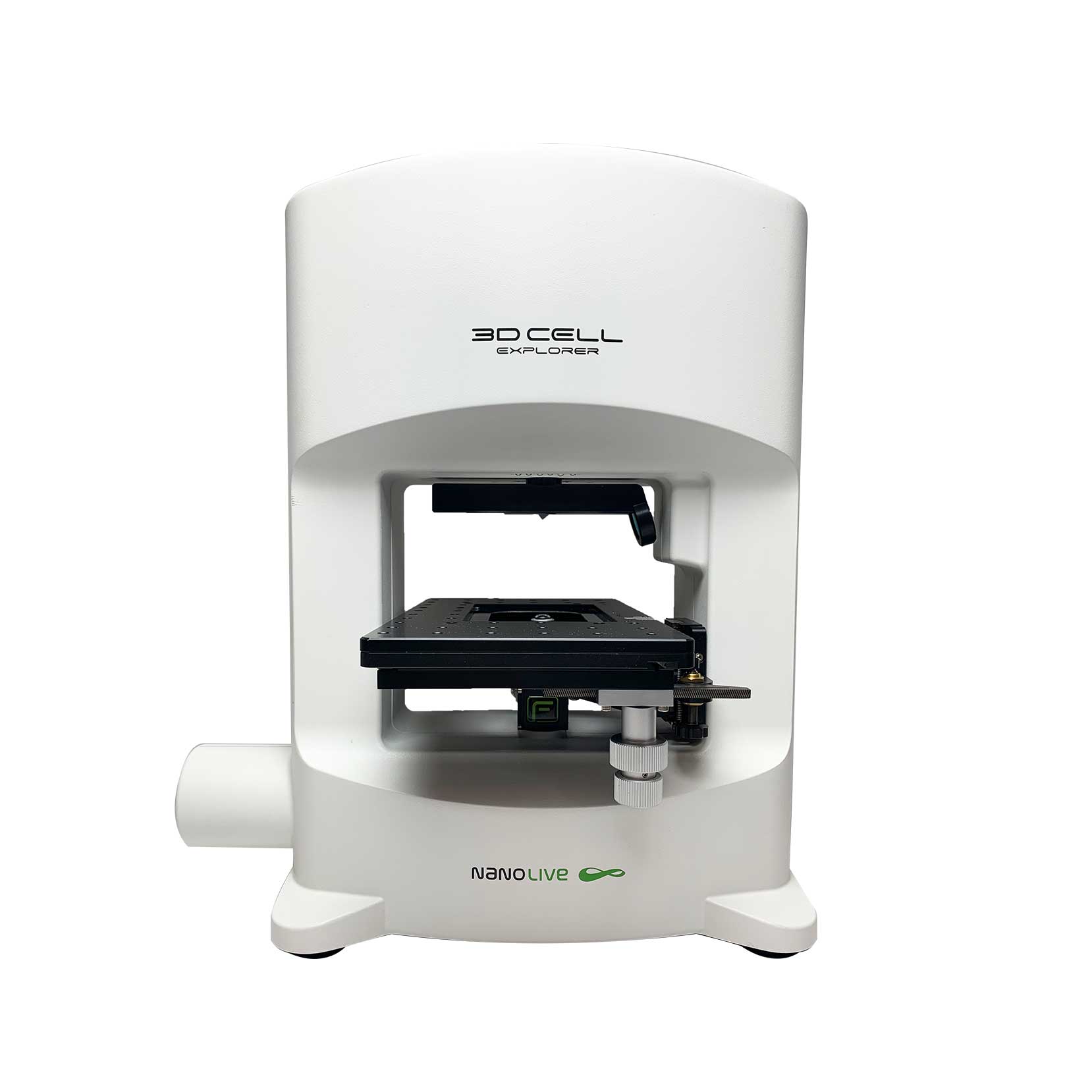
3D CELL EXPLORER-fluo
Multimodal Complete Solution: combine high quality non-invasive 4D live cell imaging with fluorescence
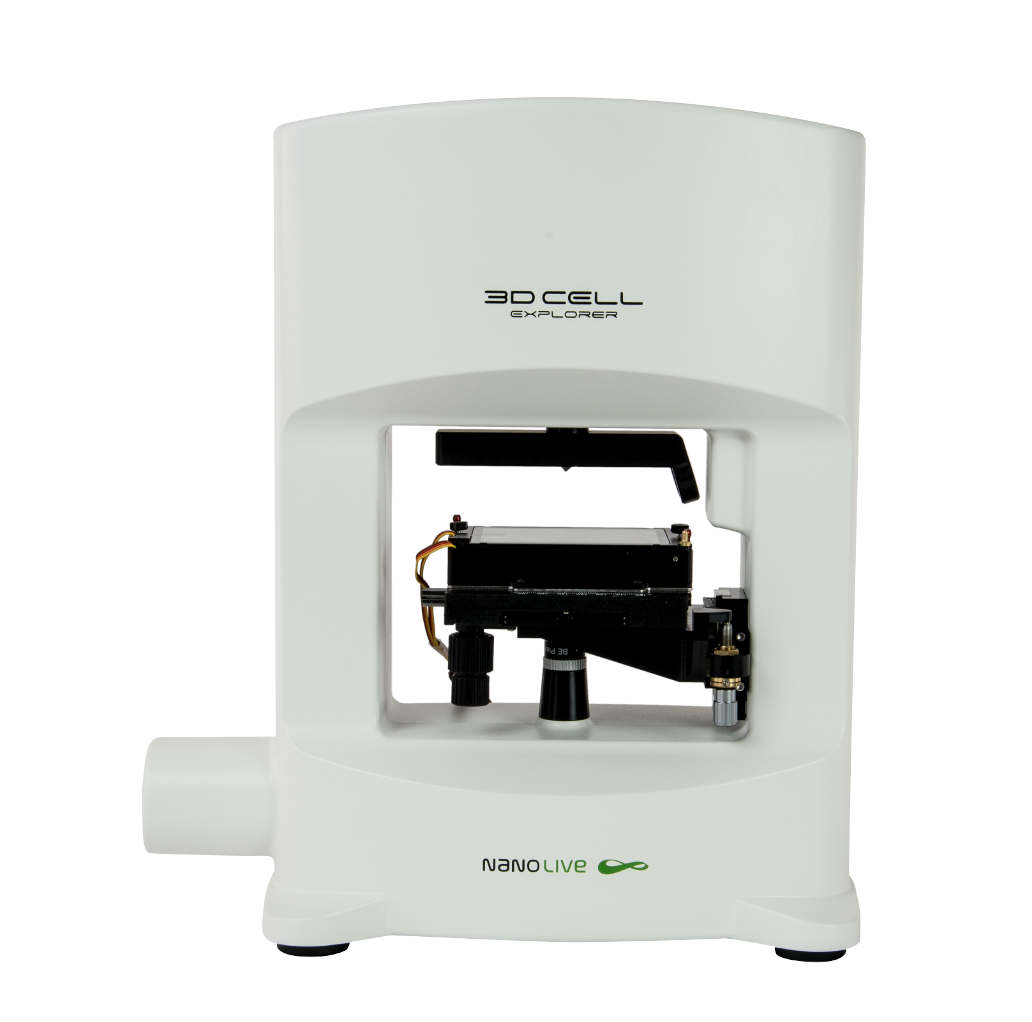
3D CELL EXPLORER
Budget-friendly, easy-to-use, compact solution for high quality non-invasive 4D live cell imaging

