Endocytosis
Endocytosis refers to the process of internalization of substances into the cell.
There are two main types of endocytosis: phagocytosis and pinocytosis. While during phagocytosis, large particles and bacteria are engulfed, in pinocytosis fluid and molecules contained in it are brought into the cell.
Endocytosis involves cytoskeletal and structural modifications. The 3D Cell Explorer allows visualizing of fine membrane deformations and posterior vesicle formation that occur in endocytic processes.
Phagocytosis: Marker-free Imaging of Cryopreserved Human M1 Macrophages
In this video – obtained with Nanolive’s 3D Cell Explorer – we present cryopreserved human M1 macrophages from PromoCell in cell culture (video 1). The 3D Cell Explorer allows to image these living macrophages in a novel, marker-free fashion. A special note goes to the visualization of membrane ruffling as waves arising at the leading edge of lamellipodia that move centripetally toward the main cell body. Macrophages were imaged for over 24h at a frequency of 1 image every 10 seconds.
Phagocytosis: The Perfect Murder – Macrophage Cell Killed by T-cells
In this movie, we can observe macrophages and T-cells interacting. Naive T-cells are being presented with antigens by the macrophages which “instruct” T-cells on what type of cells to target (such as cancer cells) and kill. During this interaction, T-cells can play a role in immune system homeostasis by killing the macrophage presenting the antigen.
Information on Z axis (depth) was processed so that a color scale (a gradient of color ranging from blue to pink) was applied to it, providing a sense of spacial organization in that axis. Full cells or cell components closer to the dish surface were colored blue, while pink accounted for cells or cellular content positioned further from the dish surface.
Cell drinking: a closer look on macropinocytosis
In this video we can observe CHO cells carrying out pinocytosis. The membrane extends forming arm-like claws that reconnect resulting in a macropinosome. Once at the cytoplasm, the macropinosome fuses with a lysosome.
Scientific Publication
Endocytosis-mediated mitochondrial transplantation: Transferring normal human astrocytic mitochondria into glioma cells rescues aerobic respiration and enhances radiosensitivity
This is a publication in the journal Theranostics from users of the Nanolive’s 3D Cell Explorer in the Laboratory of Heavy Ion Radiation Medicine of Chinese Academy of Sciences in Lanzhou, China.
Mitochondrial metabolic abnormalities have been linked to resistance to radiotherapy due to radiation cytotoxicity in cancer cells[1]. In aerobic conditions, an increased glycolysis and lactate production instead of the much more efficient oxidative phosphorylation are observed in cancer cells[2]. This change in metabolism is known as the Warburg effect[2].
Sun and colleagues have studied the radiosensitization effects induced by transplantation of mitochondria from normal human astrocytic into glioma cells. The focus of their study was to identify the mechanism of free mitochondrial transfer into host cells via a NAD+-CD38-cADPR-Ca2+-endocytosis pathway. Their results show that starvation treatment led to a decreased Warburg effect and a recuperation of aerobic respiration. Mitochondrial transplantation into glioma cells could reduce resistance to radiotherapy.
The 3D Cell Explorer was used to obtain live images of the dynamic behaviour of endocytosis during starvation treatment and of the interaction between this process and mitochondria. Nanolive imaging has proven to be a method of choice in organelle dynamics research, due to its non-invasive and phototoxicity free imaging. Their full publication is available here!
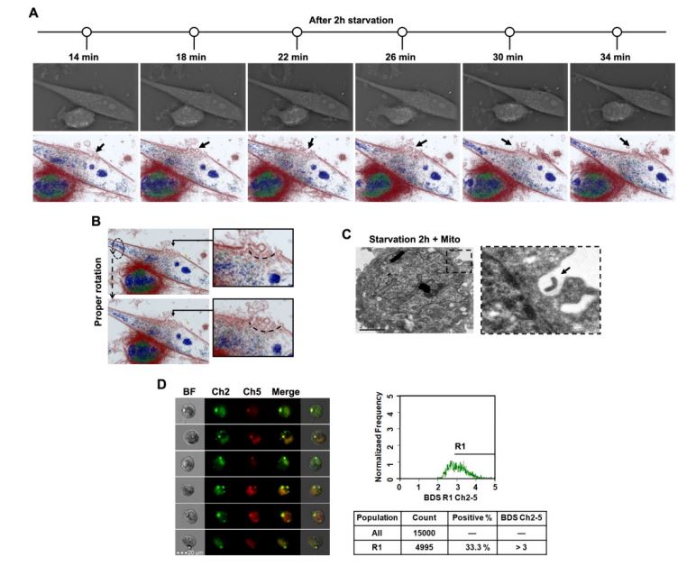
Figure: Transplantation of isolated mitochondria into U87 cell through endocytosis
[1] C. Sun et al., “Endocytosis-mediated mitochondrial transplantation: Transferring normal human astrocytic mitochondria into glioma cells rescues aerobic respiration and enhances radiosensitivity,” Theranostics, vol. 9, p. 12, 2019.
[2] O. Warburg, F. Wind, and E. Negelein, “THE METABOLISM OF TUMORS IN THE BODY.,” J. Gen. Physiol., vol. 8, no. 6, pp. 519–30, Mar. 1927.
Video Library
Live Human M1 Macrophages imaged for 24h. E.coli being engulfed through phagocytosis
Live Human M1 Macrophages imaged for 4h. E.coli being engulfed through phagocytosis