Virology
Virology is the scientific discipline targeting the study of viruses and viral diseases.
Nanolive imaging allows for the observation of processes of infection and exploitation of host cells for virus reproduction in real time and in a 3D fashion.
Interactions between the host organism physiology and immune system as a response of viral diseases can also be monitored, and 3D localization of virus inside living cells is possible.
Virology: Adenovirus infection in osteosarcoma cells
In this footage, obtained using Nanolive’s 3D Cell Explorer-fluo we observe the progression of GFP-labelled adenovirus in human bone osteosarcoma epithelial (U2OS) cells. Refractive images were taken every 15 s for 25 mins, and fluorescence images were taken every 5 mins using the FITC channel.
Special thanks to Prof. Clodagh O’Shea from the Salk Institute for Biological Studies (La Jolla, CA, USA).
Virology: Human papillomavirus infection in HeLa cells
In this footage, obtained using Nanolive’s 3D Cell Explorer-fluo, cervical cancer cells (HeLa cell line) were infected with HPV labelled with Cy5 and the internalization process was followed over time. One image was obtained every 5 mins for 5 h 20 min.
Special thanks to Prof. Wilbe Martin Kast, from the Kast Lab in Los Angeles (California, USA) for preparing the samples.
Virology: Virus-induced cytopathic effects in living cells
This video reveals virus-induced cytopathic effects in living cells. Virus infections have a physical signature (Refractive Index) which can be detected with the 3D Cell Explorer.
The Greber Group of the University of Zurich published a paper describing how Nanolive imaging reveals distinct patterns of virus infections in live cells with minimal perturbation.
Read the author’s exclusive interview on how different viruses infect living cells and discover more videos:
Compare Nanolive's microscopes

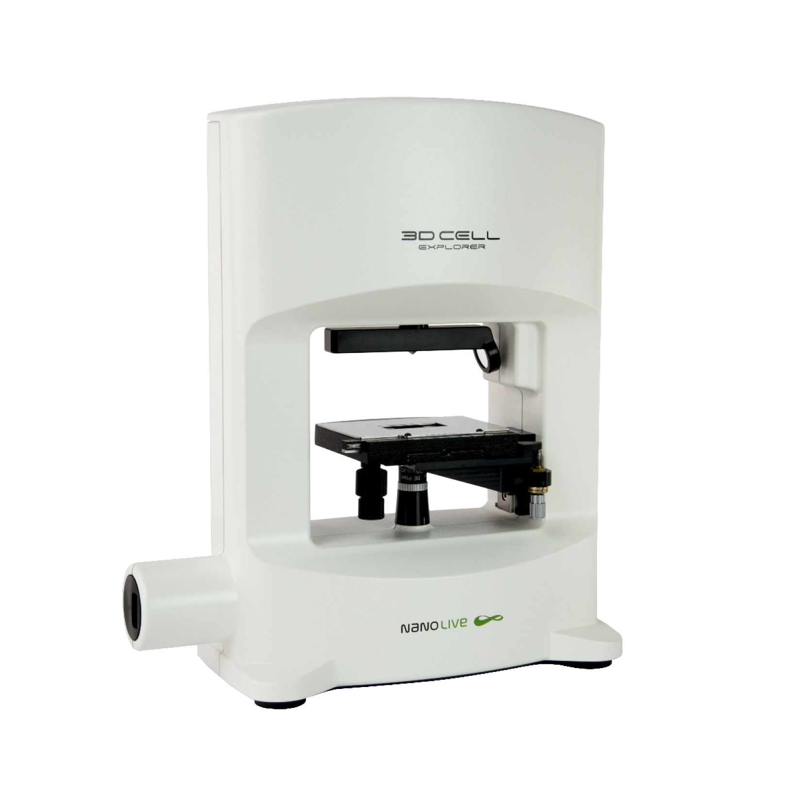
3D CELL EXPLORER
Budget-friendly, easy-to-use, compact solution for high quality non-invasive 4D live cell imaging

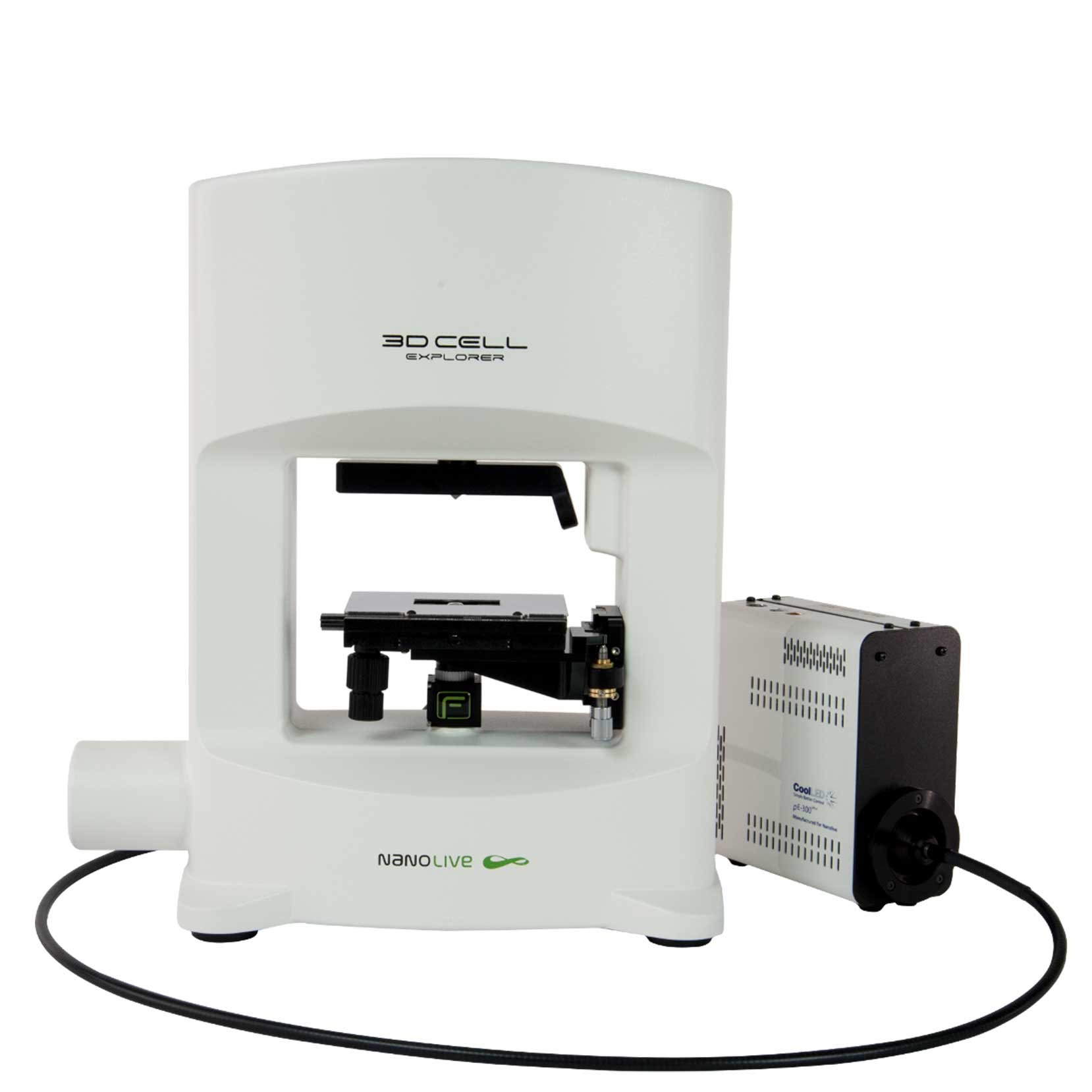
3D CELL EXPLORER-fluo
Multimodal Complete Solution: combine high quality non-invasive 4D live cell imaging with fluorescence

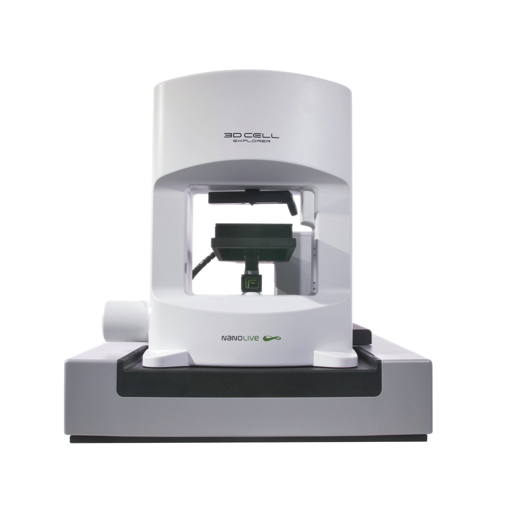
CX-A
Automated live cell imaging: a unique walk-away solution for long-term live cell imaging of single cells and cell populations
